
the Creative Commons Attribution 4.0 License.
the Creative Commons Attribution 4.0 License.
 | 28 Jul 2026
Ice-nucleating particles in Greenlandic glacial outwash plains
Nora Bergner
Grace Marsh
Kevin Barry
Larissa Lacher
Alexander Böhmländer
Joanna Alden
Carina Ahlqvist
Ianina Altshuler
Lisa Bröder
Daniel Farinotti
Lionel Favre
Coline Guillosson
Benjamin Heutte
Kristina Höhler
Roman Pohorsky
Julian Weng
High-latitude dust (HLD) represents a source of ice-nucleating particles (INPs) with potential impacts on cloud formation and radiative forcing in the Arctic. Previous studies have shown that HLD can exhibit high ice-nucleating activity at high subzero temperatures, likely linked to a biological component. Yet, comprehensive assessments of HLD ice-nucleating characteristics and sources remain limited, especially in Greenland. Here, we show that glacial dust from three outwash plains in southwestern Greenland effectively nucleates ice at temperatures relevant for mixed-phase clouds, but with lower ice-nucleating activity than other HLD regions. Ice-nucleating activity of glacial dust shows high variability and is largely driven by small amounts of organic and biological material, as indicated by sample treatments and positive correlations of ice-active site densities per mass with total organic carbon and microbial abundance. Dust from the Narsarsuaq glacial outwash plain likely acted as a localized rather than a regional INP source during summer, as indicated by higher atmospheric INP concentrations above −20 °C and similarities between atmospheric and bulk dust INP spectra at the outwash plain site compared to other sites in the region. The atmospheric INP population was generally dominated by primarily biological and other organic contributions, highlighting the importance of better constraining biogenic INP emissions in the Arctic. Overall, the ice-nucleating activity of glacial dust in southwestern Greenland lies within the lower range of reported HLD INP activity, suggesting that highly active HLD parameterizations may overestimate INP concentrations in this region. This highlights the importance of region-specific INP parameterizations for improving representation of cloud processes and climate impacts in the Arctic.
- Article
(11773 KB) - Full-text XML
-
Supplement
(3988 KB) - BibTeX
- EndNote


Climate-relevant properties of mid- and high latitude mixed-phase clouds are sensitive to the aerosol population, particularly ice-nucleating particles (INPs) (e.g., Storelvmo, 2017; Tan et al., 2016; Vergara-Temprado et al., 2018). INPs initiate heterogeneous ice-formation within the temperature range from 0 to −38 °C and can also aid ice formation below −38 °C (Hoose and Möhler, 2012; Kanji et al., 2017; Murray et al., 2012). In mixed-phase clouds, INPs influence the partitioning between cloud liquid and ice water content through both the Wegener-Bergeron-Findeisen mechanism (Korolev, 2007) and secondary ice-production pathways (Field et al., 2017; Korolev and Leisner, 2020). These microphysical processes ultimately impact the radiative properties and lifetime of clouds (Ceppi et al., 2017; Storelvmo et al., 2015). The cloud-phase feedback, where changes in cloud phase under a warming climate alter the Earth's radiation budget, thus strongly depends on the current and future abundance and characteristics of INPs (Murray et al., 2021). However, identification of INP sources, quantification of INP concentrations, and understanding of their freezing characteristics and variability remain incomplete, particularly in high-latitude environments.
Globally, the dominant source of INPs at relatively low temperatures (approximately < −20 °C) is mineral dust emitted from arid and semi-arid regions (Atkinson et al., 2013; Boose et al., 2016; DeMott et al., 2003; Sassen et al., 2003). In the Arctic, long-range transported mineral dust mainly originates from Asia and North Africa (Huang et al., 2015; Kawai and Matsui, 2025). The strongest influence occurs during winter and spring and at higher altitudes (Groot Zwaaftink et al., 2016; Kawai et al., 2023; Shi et al., 2022). More recently, increasing attention has been directed toward high-latitude dust (HLD), defined as dust from locations north of 50° N or south of 40° S (Bullard et al., 2016; Meinander et al., 2022), which may act as a regional source of INPs in polar regions (Barr et al., 2023; Kawai et al., 2023; Paramonov et al., 2018; Sanchez-Marroquin et al., 2020; Shi et al., 2022; Tobo et al., 2019; Xi et al., 2022). HLD sources are commonly linked to glacial and periglacial environments, particularly glacial outwash plains (Bullard et al., 2016). Emissions often peak in autumn due to abundant sediment supply and higher wind speeds (Bullard et al., 2023; Crusius et al., 2011). In addition to transported and local mineral dust, primary biological aerosol particles (PBAPs) have been identified as INPs in Arctic environments. These include bacteria, pollen, plant debris, fungal spores, and viruses from both marine and terrestrial environments, and are particularly active during summer and at relatively high subzero temperatures (Beck et al., 2024; Pereira Freitas et al., 2023; Šantl-Temkiv et al., 2019; Wieber et al., 2025). Beyond PBAPs, organic material such as extracellular polymeric substances, polysaccharides, humic-like substances, proteins, and other biogenic macromolecules can act as INPs (Dreischmeier et al., 2017; O'Sullivan et al., 2015; Pummer et al., 2015). Both intact PBAPs and organic macromolecules can attach to and be transported with mineral dust, thereby enhancing its ice-nucleating activity (Conen et al., 2011; O'Sullivan et al., 2014; Prospero et al., 2005), a mechanism that likely contributes to the ice-nucleating activity of HLD (Barr et al., 2023; Tobo et al., 2019; Xi et al., 2022).
Tobo et al. (2019) reported remarkably high ice-active site densities per mass at temperatures between −5 and −25 °C from dust collected in an outwash plain in Svalbard, likely linked to small amounts of organic matter that may be more ice-active in cold than temperate or warm climates. Other HLD studies also document high ice-nucleating activity at relatively high temperatures, though with notable variability. For example, Xi et al. (2022) observed dust near the Kaskawulsh Glacier in Yukon, Canada, with ice-active site densities per mass (nm, the cumulative number of ice-active sites per gram of dust) about two orders of magnitude lower than that of the Svalbard samples, showing biological contributions above −15 °C but mineral dominance at lower temperatures. Airborne dust from the Copper River Delta in Alaska exhibited high ice-active site densities per surface area (ns, cumulative number of ice-active sites per unit surface area of dust), with heat-sensitive INPs active down to −25 °C (Barr et al., 2023). Barr et al. (2023) further suggested that watershed characteristics, including the presence of vegetation and forest as sources of organic and biological INPs, may explain differences among regions. In contrast, ice-nucleating activity reported from Iceland, an active HLD source (Dagsson-Waldhauserova et al., 2013), is more comparable to low latitude dust sources (Paramonov et al., 2018; Sanchez-Marroquin et al., 2020). Modelling studies show that HLD contributes substantially to the dust load and INP population of the lower troposphere in summer and autumn (Groot Zwaaftink et al., 2016; Kawai et al., 2023; Shi et al., 2022). Incorporating a parameterization based on the high ice-nucleating activity of Svalbard HLD yields more than 100 times higher INP concentrations in the Arctic lower troposphere during these seasons and better reproduces observations (Kawai et al., 2023). With climate change, increasing HLD emissions with high ice-nucleating activity may partially offset the reduced INP activation expected at higher temperatures (Matsui et al., 2024). Taken together, these sensitivities in combination with the scarcity and heterogeneity of available observations highlight the need to better understand the ice-nucleating properties of different HLD sources.
Greenlandic glacial outwash plains have been identified as significant dust sources (Bullard et al., 2023; Bullard and Mockford, 2018), but their INP characteristics remain poorly constrained. Regional terrestrial environments in Greenland, including biogenic material, are known to contribute to INPs in summer (Šantl-Temkiv et al., 2019; Sze et al., 2023; Wex et al., 2019), yet the specific extent and properties of Greenlandic dust have yet to be determined. While Greenland accounts for only a small fraction of the total Arctic dust load (∼ 0.1 % north of 60° N; Groot Zwaaftink et al., 2016), the dust could exert a notable local to regional influence. Estimates show that up to 67 % of deposited dust on the ice sheet originates from Greenlandic sources (Groot Zwaaftink et al., 2016). Greenlandic outwash plains are expected to expand and proglacial areas have undergone pronounced changes in the last decades (Grimes et al., 2024). These changes include increased availability of fine sediments and expanding vegetation cover. Both processes may influence INP emissions, highlighting the variable and evolving nature of Greenlandic INP sources.
To address the uncertainties regarding Greenlandic dust sources as INPs, we use field and laboratory measurements to (i) quantify the ice-nucleating activity and variability of dust in glacial outwash plains in southwestern Greenland, (ii) identify sources (primary biological, other organic, mineral) contributing to the ice-nucleating activity, and (iii) assess atmospheric INP concentrations in southern Greenland and their possible origins, including HLD.
2.1 Measurement overview
Field measurements were conducted in southern and southwestern Greenland during 2023 and 2024 as part of the projects “Greenlandic Fjord Ecosystems in a Changing Climate: Socio-cultural and Environmental Interactions” (GreenFjord) and “Ecological and Climate Impacts of Greenlandic Glacial Outwash Plains” (ECO-Plains). The primary field campaign occurred from 16 June to 5 August 2023 in Narsarsuaq and Narsaq, focusing on atmospheric measurements and dust sampling. Additional dust samples were collected in Igaliku (May 2023) and Kangerlussuaq (July 2023 and July 2024), as well as in Narsarsuaq in July 2024 (Fig. 1).

Figure 1Field work sites overview. (a) Sample locations in Kangerlussuaq, (b) overview of sampling regions in Greenland, (c) main study area in Southern Greenland with zoomed-in views of (d) Narsaq with the atmospheric stations at Narsaq International Research station (NIRS) and hill, Narsarsuaq including the plain and col atmospheric stations, and (f) Igaliku. Map background imagery was obtained from the Greenlandic satellite orthophoto mosaic provided by Dataforsyningen and accessed via QGreenland (Moon et al., 2023).
Narsarsuaq, Narsaq, and Igaliku are situated in the fjords of southern Greenland, with the Narsarsuaq outwash plain stretching in front of the glacier Kiattuut sermiat, while Igaliku is located at the Aniaaq fjord, near the glacier Jespersen Brae. Narsaq lies at the intersection of Tunulliarfik fjord and Ikersuaq fjord. The Kangerlussuaq outwash plain is located on the southwestern coast, spanning from Russell Glacier to the head of Kangerlussuaq fjord. These glacial outwash plains exhibit varying vegetation covers, including unvegetated areas and regions with sparse, low-level vegetation such as grasses, mosses, lichens, and, in some cases, lower shrub vegetation. Shrub vegetation is dominant around Narsaq.
2.1.1 Bulk dust samples
Dust samples were collected following a chronosequence approach (Bradley et al., 2016), where locations farther from the glacier have been exposed longer to the atmosphere and weathering processes following glacial retreat. These sites may show advanced microbial succession or soil development, potentially influencing the dust's ice-nucleating activity. We distinguish between two types of dust samples: transect and microbiology. Transect samples were collected along glacier-to-fjord transects, targeting surface sediments (top ∼ 1 cm) from primarily vegetation-free areas or river deposits, where dust emissions are likely. Samples were air-dried under a laminar flow hood and sieved to < 45 µm to focus on the particle size fraction most relevant for atmospheric transport, following common approaches in similar studies (e.g., sieving to sizes between 32–63 µm in Barr et al., 2023; Boose et al., 2016; Hamzehpour et al., 2022). Size distributions of the transect samples, measured by a laser diffraction particle size analyzer (0.017–2000 µm, LS 13 320, Beckman Coulter, USA), show that particles are below 10 µm with a main mode below 0.1 µm for most samples (Fig. S1 in the Supplement). Microbiology samples (top ∼ 5 cm) were collected at two sites each in Narsarsuaq and Kangerlussuaq, with site 1 closer to the glacier than site 2. Each site included multiple subsites from both vegetated and unvegetated surfaces. Since these samples were primarily analyzed for microbiological purposes, they were air-dried but not sieved, allowing for better comparison between microbiological measurements and INP analyses for this sample type. Although transect and microbiology samples were collected from different depth intervals (top ∼ 1 cm and top ∼ 5 cm, respectively), analysis of microbial community composition from the same sites found no significant differences in ice-nucleating genera across these soil depths (Marsh et al., 2026), suggesting that this difference does not affect the interpretation of the results. Details and sampling locations are shown in Table S1 and Fig. 1, with photos of some sampling locations in Fig. S2.
Additional bulk dust samples were collected for comparison in three glacial outwash plains in the Swiss Alps, allowing us to assess whether ice-nucleating properties observed in Arctic samples are specific to Arctic conditions or represent a more general feature of glacially derived dust, including at mid-latitudes. The sampling sites are shown in Table S4 and Fig. S23.
2.1.2 Atmospheric measurements
Atmospheric measurements were conducted in both Narsarsuaq and Narsaq with two sites per location (Fig. 1), each equipped with small solar- and battery-powered stations. In Narsarsuaq, one station was installed on the outwash plain, hereafter referred to as the plain station. A second station was positioned on a small col, located approximately 1.1 km northeast of the plain station at an elevation of 300 m a.s.l., hereafter referred to as the col station. In Narsaq, measurements were performed at the Narsaq International Research Station (NIRS), where a more extensive suite of instruments was deployed, the set-up is described in detail by Alden et al. (2026). The second Narsaq site was situated on Tasigaaq hill (hereafter referred to as hill station), approximately 1.6 km northeast of NIRS at an elevation of 372 m a.s.l. Photos of all measurement stations are provided in Fig. S2.
At each site, meteorological variables including atmospheric pressure, temperature, relative humidity, solar radiation, wind/gust speed and precipitation were measured with ATMOS41 GEN 2 weather stations (METER Group, USA). Particle number concentrations and size distributions were obtained with optical particle counters: at NIRS, in the size range 0.19–93.06 µm (optical diameter, dopt), using a Fidas Frog (Palas, Germany), and at the plain, col, and hill sites in the size range 186–3370 nm (dopt) using a Portable Optical Particle Sizer (POPS, Handix Scientific, USA). The reported size ranges exclude the smallest size bins due to higher measurement uncertainty (Mei et al., 2020). Aerosol filter samples were collected for INP measurements using a custom-built aerosol filter sampler. The system consisted of a stainless-steel inlet, an in-line plastic filter holder (Whatman®, Cytiva, USA), a mass flow controller (Sensirion AG, Switzerland), and a pump (KNF Neuberger GmbH, Germany). Inlet heights were 100 cm above ground at the plain station, 116 cm at the col station, 3 m at NIRS, and 142 cm at the hill station. Nuclepore™ polycarbonate filters (47 mm diameter, 0.2 µm pore diameter) were prepared according to the protocol described by Barry et al. (2021) and inserted into the holders under clean conditions. Filter samplers were operated at a flow between 5 and 10 std L min−1 (std denotes volumetric flow referenced to standard temperature and pressure) for a typical duration of 1–3 d, with some filters up to 14 d due to logistical reasons. An overview of all filters analyzed for INPs is provided in Table S2.
2.2 Ice-nucleation freezing measurements
Bulk dust and filter samples were analyzed for INPs using the Sion Particle Ice Crystallization Experiment (SPICE), an immersion-mode INP measurement set-up described in detail in Appendix A. All samples were stored frozen during the campaign (approximately −20 °C) and transported frozen to the laboratory (< 0 °C). Filter samples remained frozen until INP analysis (approximately −20 °C), whereas transect dust samples were dried and sieved at room temperature shortly after the campaign and then stored frozen (approximately −20 °C) until INP experiments were performed. Storage and freeze-thaw cycles can affect ice nucleation activity of both mineral and biological samples, typically causing INP degradation (Beall et al., 2020; Perkins et al., 2020; Wex et al., 2015), although the effects are not fully understood. Frozen storage was chosen to minimize potential INP degradation.
Suspensions were made in pre-rinsed 50 mL Corning® polypropylene centrifuge tubes (Cat. No. 10788561, Corning Inc., USA) with molecular-biology-free reagent water (Cat. No. W4502, Sigma Aldrich, USA; hereafter referred to as SA water). Initial suspensions were prepared at approximately 2 g L−1 by weighing a defined mass of dust (e.g., 40 mg) and adding SA water (e.g., 20 mL). Four sequential 10-fold dilutions were then prepared for bulk dust samples (0.5 mL sample, 4.5 mL SA water), resulting in a concentration range of approximately 2 to 0.0002 g L−1. The dust samples formed homogeneous suspensions in water, although some settling was observed in the sample tube with the highest dust concentration during preparation, which was addressed by repeated shaking. Filter samples were suspended in 12 mL of SA water (8 mL for procedural blank filters, as these were processed earlier in the study before the protocol was optimized), and the aerosol was washed off by placing the Corning tube in a tube rotator at 60 rpm for 20 min. Two 15-fold dilutions (0.4 mL sample and 5.6 mL SA water) were prepared for the filter samples. The undiluted suspensions and corresponding dilutions were pipetted in 50 µL droplets into the wells of two single use PCR-trays (Cat. No. 781368, Brand, Germany). For filter samples, the 192 wells were distributed as follows: 40 wells for the initial suspension, 56 wells for the 15-fold dilution, 64 wells for the 225-fold dilution, and 32 wells for background SA water. For bulk samples, the wells were distributed in blocks of 32 for the initial suspension, for each dilution, and for the background. Transparent foil (Cat. No. AXYPCR-TS, Axygen, Corning Inc., USA) was placed on top of the PCR trays to prevent contamination. The PCR trays were placed into the two aluminum cooling blocks of SPICE and cooled down with a chiller (RP 245 E, Lauda, Germany) from 2 °C to approx. −35 °C at a cooling rate of −0.33 °C min−1. Particle settling during the freezing experiment is a potential concern for bulk dust samples containing coarser particles. However, size distributions of the sieved transect samples show that particles are predominantly in the fine mode with a main mode below 0.1 µm (Fig. S1), making settling unlikely for this sample type. The unsieved microbiology samples follow the same methodology as Barry et al. (2023), enabling direct comparison with their dataset, but settling of larger particles cannot be excluded for this sample type, introducing additional uncertainty to the reported INP concentrations.
From the observed freezing events, the ice nucleation active sites per unit volume of water as a function of temperature, INPsus,corr(T), also referred to as cumulative INP spectrum, were calculated following Vali (1971, 2019), including a correction for the water background (Eq. A5). The ice-active site densities per mass (nm) and per surface area (ns) were calculated with equations A7 and A8, respectively. The specific surface area of the transect samples was estimated geometrically from laser diffraction particle size distributions (LS 13 320, Beckman Coulter, USA), assuming spherical particles with a density of 2650 kg m−3, using only size bins below 45 µm consistent with the sieving cutoff applied prior to measurement (, where fi is the normalized volume fraction and di the bin centre diameter). The resulting geometric specific surface area values are provided in Table S1. Atmospheric INP concentrations (NINP) were computed from the ratio of washing water to sampled air volume (Eq. A6). The minimum detectable atmospheric INP concentration, defined as the concentration corresponding to one frozen well given the assay parameters and sampled air volume, ranges from 4.5 × 10⁻⁵ to 9.9 × 10⁻⁴ L−1 across samples depending on the sampled air volume. Absent data points at higher temperatures are consistent with INP concentrations falling below this limit. 95 % confidence intervals for all INP spectra were calculated according to Eq. (2) in Agresti and Coull (1998) and more detailed data processing and quality checks are described in Sect. A1.3. Procedural filter blanks at each station were taken by inserting the filter holder with a filter but without turning on the air flow. The background filters showed lower frozen fraction curves compared to the filter samples (Fig. S3), but were above the typical background freezing from SA water only, with some blank filters showing non-negligible amounts of INPs. Procedural blanks were not subtracted from sample spectra and contribution is likely minor for most samples and confined to low temperatures (Fig. S4). However, background contamination introduces additional uncertainty to the reported INP concentrations, particularly for samples with low INP concentrations.
For compositional information, we performed heat and hydrogen peroxide treatments to a subset of bulk dust and filter samples. Heat treatment denatures proteins, with proteinaceous INPs in different biological materials (e.g., bacteria, fungal spores, lichen) found to deactivate between 40–60 °C (Fröhlich-Nowoisky et al., 2016; Hara et al., 2016; Kieft and Ruscetti, 1990; Pouleur et al., 1992). The difference between untreated and heat treated spectra is therefore often used as an indicator of proteinaceous, likely biological INPs (e.g., Conen et al., 2011; Hill et al., 2016; O'Sullivan et al., 2014; Pouleur et al., 1992). Hydrogen peroxide treatment oxidizes organic matter, leaving spectra that reflect the remaining inorganic or mineral INPs (e.g., Conen et al., 2011; Hill et al., 2016; McCluskey et al., 2018a). While these treatments are useful indicators for the possible origin of INPs, they have limitations and are not entirely selective. Heat treatment can reduce the ice-nucleating activity of certain minerals, including quartz and plagioclase feldspar (Daily et al., 2022), while some thermostable biological material can remain active (Pummer et al., 2012). Similarly, hydrogen peroxide treatment can affect carbonate minerals, although other mineral components are likely unaffected (Hill et al., 2016; O'Sullivan et al., 2014; Tobo et al., 2019). For the heat treatment, a Corning tube containing 3 mL of the sample suspension was placed into a boiling water bath (98.4 °C) for 20 min. For the hydrogen peroxide treatment, 1 mL of 30 % H2O2 (Cat. No. 216763-100ML, Merck Millipore, Germany) was added to 2 mL of the sample suspension and placed into a boiling water bath with UV light for 20 min. The UV radiation generates highly reactive hydroxyl radicals that oxidize organic compounds. Remaining peroxide was neutralized by adding small quantities (90 to 130 µL) of 0.1 µm filtered catalase (Cat. No. MPB-210042910-10ML, MP Biomedicals, USA) to avoid freezing point depression. Dilution series and freezing experiment for the treated samples were performed as described above.
In total, we analyzed 32 bulk dust samples with 28 heat and 5 hydrogen peroxide treatments, as well as 38 filter samples with 5 heat and 5 peroxide treatments (Table S1). In addition, 14 procedural blank filter samples were analyzed. Additional experiments including SA blanks and validation material for the SPICE characterization are shown in Sect. A2.2 and A2.3.
2.3 Ancillary measurements
To evaluate the role of organic matter for ice-nucleating activity, total organic carbon (TOC) was measured for all dust samples using catalytic high-temperature combustion with CO2 quantification via non-dispersive infrared detection (Elementar Enviro TOC analyzer, Elementar Analysensysteme GmbH, Germany). The microbial abundance in the microbiology dust samples was assessed using two different methods: flow cytometry and plate culturing. For flow cytometry, bacterial cells were extracted from 1 g soil suspended in 9 mL 1 × phosphate-buffered saline (PBS) (sterile filtered at 0.2 µm), following a protocol adapted from Górniak et al. (2017). After shaking (120 rpm, 2 h, 10 °C) and fixation with 3.7 % formaldehyde, cells were separated from soil particles via Histodenz (Cat. No. D2158, Sigma-Aldrich, USA) density centrifugation (17 000 × g (relative centrifugal force), 90 min, 4 °C) (Frossard et al., 2016). The upper aqueous phase was extracted, stained with SYTO13 (50 µM), and analyzed using a NovaCyte flow cytometer (ACEA, Bioscience, USA) (flow rate 14 µL min−1), with cell counts determined from forward and side scatter signals. For plate culturing, 1 g of soil was suspended in 9 mL 1 × sterile PBS, shaken for 2 h at 10 °C, and allowed to settle for 15 min. Serial dilutions were plated (500 µL) onto R2A agar media (Neogen Culture Media, USA) and incubated at 12.5 °C (the average temperature across sites). Colony-forming units (CFUs) were counted after three weeks.
X-ray diffraction (XRD) measurements were performed on a subset of representative samples of each outwash plain to determine the relative abundance of ice-nucleating active minerals. Diffractograms were collected using a D8 Discover Vario (Bruker, USA) with Cu Kα1 radiation, and mineral phases were identified using the Rietveld refinement method. Semi-quantitative estimates of relevant mineral abundances are summarized in Table S3.
2.4 Principal component analysis
To examine similarities between bulk dust and aerosol INP spectra, we performed a principal component analysis (PCA) on derived spectral features, specifically slopes and logarithmic concentration ratios, following the approach of Barry et al. (2023). Deriving spectral features enables comparison across sample types measured in different units (nm vs. NINP), since slopes and logarithmic concentration ratios describe the shape of the INP spectra rather than absolute magnitudes. Slopes characterize the temperature-dependent steepness while logarithmic ratios capture relative concentrations across the temperature range, and both features can be indicators of the underlying INP source (e.g., Barry et al., 2023; Sze et al., 2023). For this analysis, INP concentrations between −8 and −20 °C were log-transformed (log10) and missing values were interpolated or extrapolated linearly. From these spectra, we derived (i) slopes in 2 °C windows shifted in 1 °C steps, and (ii) logarithmic ratios of mean INP concentrations in each 2 °C window relative to −15 °C. The 2 °C window size balances spectral resolution against measurement noise, and the 1 °C step size ensures continuous coverage across the temperature range. −15 °C was selected as a reference temperature as it falls near the center of the analysis range and is a commonly used benchmark temperature for INP comparisons relevant for mixed phase clouds (Barry et al., 2023; Morrison et al., 2012), although results are not sensitive to this choice. The resulting feature set (22 variables across 70 samples) was standardized (z-scores) prior to PCA, which was applied to the combined dataset of bulk and aerosol samples.
3.1 Ice-nucleating properties of dust in southwestern Greenlandic glacial outwash plains
The following sections present an analysis of the ice-nucleating properties of dust collected from glacial outwash plains in southwestern Greenland. We first characterize the ice-nucleating activity of bulk dust samples across different sites and how they compare to other high-latitude dust sources in Sect. 3.1.1. We then explore the spatial variability within the outwash plains (Sect. 3.1.2) and examine the influence of organic and biological material on the observed ice-nucleating activity through treatments (Sect. 3.1.3). Finally, we assess correlations between total organic carbon (TOC), microbial abundance, and ice-nucleating activity (Sect. 3.1.4.) to better understand the drivers of variability across samples.
3.1.1 Overview of the ice-nucleating activity of glacial dust
To characterize the bulk dust in glacial outwash plains in Greenland as a potential INP source, we present the ice-active site densities per mass (nm) and ice-active site densities per surface area (ns) in Fig. 2. The ice-nucleating activity of the glacial dust is quite variable for a given site, but similar across the three outwash plains in southwestern (Kangerlussuaq) and southern Greenland (Narsarsuaq, Igaliku), with nm values (Fig. 2a) from all sites falling within a comparable range and showing no systematic differences between locations. A subset of samples with higher ice-nucleating activity (e.g., some microbiology samples) likely indicates an influence of vegetation cover (Fig. S5). The ns values (Fig. 2c, transect samples only) are likewise similar across sites.

Figure 2Bulk dust ice-nucleating activity and comparison. (a) Ice-active site densities per mass (nm) in Narsarsuaq, Kangerlussuaq and Igaliku with the polynomial fit for all data combined () and (b) Greenlandic dust samples (black lines, same as panel (a)) compared with literature data by Tobo et al. (2019, bulk dust sieved to < 5 µm), Xi et al. (2022, dust aerosol samples with nm determined gravimetrically), Hill et al. (2016, unsieved bulk soil samples), and Barry et al. (2023, unsieved bulk sediment samples). (c) Ice-active site densities per surface area (ns,geo) of dust samples based on geometric specific surface area estimates with a polynomial fit () and (d) Greenlandic dust samples (black lines, same as panel (c)) compared with literature data by Barr et al. (2023, dust aerosol samples converted to ns,geo), Sanchez-Marroquin et al. (2020, dust aerosol samples converted to ns,geo), Xi et al. (2022, dust aerosol samples converted to ns,geo), K-Feldspar, albite, and quartz parameterizations (Harrison et al., 2019, based on ns,BET), scaled to the average abundance of the respective mineral in the sample based on XRD measurements (Table S3), Ullrich et al. (2017, parameterization based on ns,geo), Niemand et al. (2012, parameterization based on ns,geo).
The Greenlandic dust's nm values are substantially lower (sample average ∼ 2–3 orders of magnitude) compared to glacial dust collected in Svalbard (Tobo et al., 2019, < 5 µm) and midlatitude soils (Hill et al., 2016), particularly for the relatively warm temperatures (Fig. 2b). In contrast, the nm values are more comparable to airborne dust from a glacial outwash plain in Yukon, Canada (Xi et al., 2022), and sediments (not a glacial outwash plain) collected near Utqiaġvik, Alaska (Barry et al., 2023). The ns values mostly fall within the lower range of other HLD observations (Fig. 2d), including airborne dust from the Copper River Delta in Alaska (Barr et al., 2023), Icelandic dust plumes (Sanchez-Marroquin et al., 2020), and airborne dust from Yukon, Canada (Xi et al., 2022, same samples as nm). However, it should be noted that differences in sampling and normalization methods (e.g., bulk dust vs. aerosol sampling, ns based on geometric vs. BET surface area) may influence the direct comparability of these datasets.
Low latitude desert dust parameterizations (Niemand et al., 2012; Ullrich et al., 2017) fall at the upper end of the ice-nucleating activity observed in our samples but are only applicable at lower temperatures (< −12 °C). Ice-nucleating-active minerals typically control the ice-nucleating activity of desert dust samples, and we include parameterizations for potassium (K-Feldspar), albite, and quartz (Harrison et al., 2019), scaled to the range of their relative abundances determined by XRD analysis of the sieved (< 45 µm) bulk samples (Sect. 2.3, Table S3). The quartz- and albite-based parameterizations predict lower ns values than observed. While the K-Feldspar parameterization is in a similar range as the Greenland dust observations, the differences in slope suggest that other factors control the measured ice-nucleating activity.
Overall, the Greenland dust samples exhibit freezing onsets at relatively high temperatures (−6 to −10 °C) relevant for mixed-phase clouds, and show partly comparable (Xi et al., 2022), but generally lower ice-nucleating activity than other HLD sites (Tobo et al., 2019; Barr et al., 2023). These findings suggest that HLD could represent a rather heterogeneous source of INPs, with variable ice-nucleation efficiencies depending on location. The variability could stem from differences in mineralogy as well as from biological and organic compounds, which have been identified as important contributors to the HLD ice-nucleating activity (Barr et al., 2023; Sanchez-Marroquin et al., 2020; Tobo et al., 2019; Xi et al., 2022). The comparable or higher K-feldspar content of the Greenland samples relative to other HLD sites (Barr et al., 2023) suggests that the lower ice-nucleating activity observed in the Greenland samples is more likely driven by differences in biological or organic contributions. In the following sections, we investigate potential sources of ice-nucleating activity and discuss possible reasons for these differences.
3.1.2 Spatial variability
To investigate potential sources of ice-nucleating activity, we first assess the spatial variability of the Greenland dust samples, depicted in Fig. 3 for nm at −15 °C, which is chosen due its relevance for mixed-phase clouds (Hanna et al., 2008; Morrison et al., 2012) and comparison to other Arctic studies (e.g., Barry et al., 2023). The ice-nucleating activity shows high spatial variability, which is largest in the Narsarsuaq glacial outwash plain. Notably, this variability does not show clear trends across the transects, suggesting that distance from the glacier, or overall position within the outwash plain does not exert a dominant influence on nm. Based on the chronosequence concept, more developed soils farther from the glacier, exposed for longer after glacial retreat, would be expected to host greater microbial abundance and biogenic material, and thus potentially higher ice-nucleating activity. However, this pattern is not observed in our samples, suggesting that local environmental factors are more likely to influence the dust's ice-nucleating activity. Moreover, the chronosequence represents a theoretical space for time substitution that is not fully applicable to glacial outwash plains, which are highly dynamic environments characterized by strong seasonal changes and episodic flooding (e.g., glacial lake outburst floods from Lake Hullet occur regularly in Narsarsuaq; Carlson et al., 2020; Carrivick and Tweed, 2019). These processes can strongly affect the presence of vegetation, organic matter, sediment deposition, and therefore also the ice-nucleating ability.

Figure 3Spatial variability of ice-nucleating activity. INP concentration per gram of bulk dust at −15 °C in (a) Kangerlussuaq, (b) Narsarsuaq, and (c) Igaliku. The red markers correspond to the transect samples that were sieved to < 45 µm. Microbiology samples refer to unsieved samples (Sect. 2.1.1). The size of the markers corresponds to the INP concentration. Map background imagery was obtained from the Greenlandic satellite orthophoto mosaic provided by Dataforsyningen and accessed via QGreenland (Moon et al., 2023).
3.1.3 Sources of ice-nucleating activity
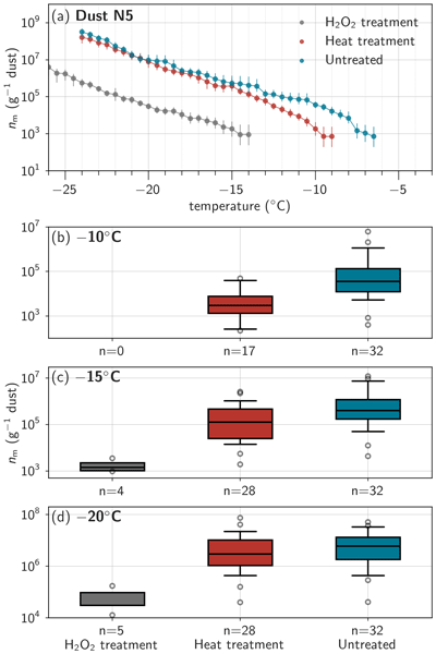
To better understand the sources of the dust's ice-nucleating activity and potential biogenic contributions, we performed heat and hydrogen peroxide treatments to the bulk dust samples, as shown in Fig. 4. The treated samples exhibit INP spectra similar to the example shown in Fig. 4a and are summarized for −10, −15, and −20 °C in Fig. 4b–d. Heat treatment reduces the ice-nucleating activity mainly at temperatures above −13.5 °C (average heat-labile fraction > 75 % for temperatures > −13 °C, n = 28) but still shows substantial contributions at −20 °C (Fig. S6). After peroxide treatment, ice-nucleating activity decreases substantially, with the remaining nm making up only a few percent (2.5 %, average across temperatures and samples, n = 5) of the nm of the untreated sample (Fig. S7).

Figure 4INP composition of dust samples. (a) nm spectra of N5, a bulk dust sample from Narsarsuaq, including heat and hydrogen peroxide treatment. Boxplots of INP concentrations at (b) −10 °C, (c) −15 °C, and (d) −20 °C of bulk dust samples without treatment (n = 32), heat treatment (n = 28), and hydrogen peroxide treatment (n = 5). Box plots show the median (center line), first and third quartiles (box edges), and whiskers extending to the 5th and 95th percentiles; outliers beyond this range are shown as individual points. Note the different y-axis in panels (b)–(d).
These treatment results suggest that organic compounds are primarily responsible for the ice-nucleating activity of the dust samples, despite the presence of ice-active minerals. While the K-feldspar parameterization falls within a similar range as the observed ice-active surface site densities (Fig. 2), the treatment results suggest a predominantly organic rather than mineral ice nucleation mechanism. However, treatments may also have non-specific effects and reduce the ice-nucleating activity of minerals (Daily et al., 2022). This organic dominance is overall consistent with the literature, where samples from various high-latitude dust sources show substantially lower ice-nucleating activity following heat and hydrogen peroxide treatment (Fig. S8; Barry et al., 2025; Tobo et al., 2019; Xi et al., 2022). When comparing with the literature, the Greenlandic samples show higher organic fractions than reported by Tobo et al. (2019) (Fig. S7), and the change in median freezing temperature (Fig. S9) between untreated and heat treated samples is similar to or even higher than in Barr et al. (2023, Fig. 3), although both studies show higher overall ice-nucleating activity (Fig. 2c, d). The larger ΔT50 values observed in our study may be influenced by differences in sample type and size (bulk dust vs. airborne particles), and indicate a higher heat-labile fraction in the Greenlandic samples. This may indicate that differences in the type or composition of organic matter play a key role in determining the magnitude of ice-nucleating activity.
3.1.4 Positive correlation of TOC, microbial abundance and INPs
Given the important contribution of organic material on the ice-nucleating activity of the dust samples, we assess the correlation between TOC and nm, as well as microbial abundance and nm (Fig. 5). Such correlations have previously been reported for water samples (Barry et al., 2023; Wilson et al., 2015), motivating a similar analysis to assess whether organic content drives INP activity in glacial dust. While TOC provides only a bulk measure of organic carbon content and cannot differentiate between specific organic compounds, it can still reveal whether the overall abundance of organic matter influences INP activity across our samples. The TOC of the samples is generally low (< 0.02–0.74 wt %, average of 0.18 wt %, 10 samples below detection limit of 0.024 wt %), and therefore all samples are lower than the 0.9 wt % measured by Tobo et al. (2019). Samples in the vicinity of vegetation show higher TOC content, but even samples with low TOC content (or just above detection limit) show important organic contributions to the dust's ice-nucleating activity (e.g., Fig. S7d). TOC significantly correlates with nm at different temperatures. The correlation coefficient shows a slight increase at lower temperatures, possibly due to reduced noise in the data, but also subsets of the data (e.g., only Kangerlussuaq or Narsarsuaq, heat treated samples) show significantly positive correlations (Fig. S10). While comparison of TOC and ice-nucleating activity of soils in different climate zones from Tobo et al. (2019) shows no simple correlation of organic content and ice-nucleating activity, the positive correlation in our samples within the same climate zone could indicate that TOC amount may contribute to the differences across HLD sites. Samples from glacial outwash plains in the Alps in Switzerland have similar or lower nm values compared to the Greenland samples (Fig. S25). They also show a significant positive correlation with TOC (Fig. S26), but generally have higher TOC values (Fig. S27). Changes in ice-nucleating activity following heat and hydrogen peroxide treatment (Figs. S28, S29) are comparable to the Greenland samples, although the Alp samples show higher inorganic fractions at temperatures < −20 °C. This supports that the ice-nucleating efficiency of TOC may differ across climatic regions (Schnell and Vali, 1973), suggesting the importance of TOC composition rather than abundance alone. However, limited information on TOC and HLD INPs are available in the literature to further investigate this relationship.

Figure 5INPs, TOC and microbial cell count. Scatterplot of (a) total organic carbon (TOC) content, microbial abundance determined by (b) colony forming units (CFU), and (c) flow cytometry (FC) vs. INP concentration per gram of bulk dust for the temperatures −10, −15, and −20 °C. For each temperature, Kendall's tau correlation coefficient (τb) and p-value are annotated.
Tobo et al. (2019) hypothesized that the ice-nucleating activity in the unvegetated glacial outwash plain in Svalbard could primarily stem from microbial sources. We find a significant correlation of microbial abundance with nm, which is consistent across temperatures when microbial abundance is assessed via colony-forming units (CFU, culturing method, Fig. 5b) and flow cytometry (FC) based cell counts (Fig 5c). The stronger correlation of nm with CFU than with FC-based total cell counts suggest that culturable, viable microorganisms are more closely linked to nm than total cell abundance. This may reflect the importance of microbial composition, with ice-nucleating microorganisms potentially being more abundant on biological surfaces than on loose outwash material (Lindow et al., 1982). Some bacterial species are highly efficient ice nucleators due to ice-binding proteins on their cell membranes, and genera known to include such species (Pseudomonas and Pseudoxanthomonas; Failor et al., 2017; Joly et al., 2013) were detected in samples from the same outwash plain (Marsh et al., 2026). However, the presence of these genera does not necessarily indicate active ice-nucleating ability, as ice-nucleating proteins may not be expressed under all environmental conditions. Ice-nucleating active species have been identified in precipitation samples at different locations including Greenland (Šantl-Temkiv et al., 2015, 2019; Stopelli et al., 2017). Notably, Šantl-Temkiv et al. (2019) found a positive correlation of airborne bacterial cell concentrations and INPs at −10 °C at Villum Research station in northern Greenland, although they did not detect the ice-nucleation active ina genes in their isolates. The positive correlation of microbial abundance and nm in our samples could directly reflect the presence of ice-nucleating bacterial species. However, TOC also correlates positively with microbial abundance, making it challenging to disentangle their relative contributions to the ice-nucleating activity. Microbiota fix carbon and nitrogen, allowing the development of more complex microbial communities, soil development and plant succession, that support more diverse ecosystems (Bradley et al., 2016; Donhauser and Frey, 2018). As microbial abundance, TOC, and nm values are higher for vegetated sites compared to unvegetated sites (Fig. S11, microbiology samples), higher microbial abundance may therefore be an indicator of an environment with more advanced soil and vegetation development, that can contain additional sources of INPs. These sources can include soil organic compounds (Hill et al., 2016; O'Sullivan et al., 2014), vegetation-derived INPs from plant surfaces that can be washed off during rainfall (Conen and Einbock, 2025; Seifried et al., 2020), and other organisms. Various fungal species have been shown to be ice-nucleating active (Fröhlich-Nowoisky et al., 2015; Huffman et al., 2013; Morris et al., 2013; Pouleur et al., 1992), and likely have an important contribution to the INP population in different Arctic environments (Barry et al., 2025; Gratzl et al., 2025; Jensen et al., 2025). Additionally, lichens (Kieft, 1988; Kieft and Ruscetti, 1990) and moss spores (Weber, 2016) have been found to be active as INPs, and could contribute to INPs at sites with biological crusts.
Overall, the ice-nucleating activity of dust in southwestern Greenlandic glacial outwash plains is lower compared to other high-latitude dust sources. The ice-nucleating activity appears to be primarily influenced by the presence of small quantities of organic and biological matter within the dust samples. Variability in nm values is closely linked to local environmental factors, including TOC content, microbial cell abundance, and presence of vegetation. Given the lower ice-nucleating efficiency compared to some other sites (Tobo et al., 2019; Barr et al., 2023), applying highly ice-efficient parameterization schemes to all HLD dust sources may overestimate atmospheric INP concentrations (Kawai et al., 2023). Moreover, while increasing Arctic vegetation (Grimes et al., 2024) may enhance ice-nucleating activity by contributing to more biogenic material, it could simultaneously reduce aeolian dust emissions, adding complexity to future projections of Arctic dust and INP dynamics. This highlights the need to further investigate biological surfaces as sources of INPs.
3.2 Atmospheric INP concentrations
Following the dust characterization as a potential INP source in the glacial outwash plains in southwestern Greenland, we assess the atmospheric INP concentrations during summer (June–August 2023) at two sites in Narsaq (NIRS and hill, fjord environment), and two sites in Narsarsuaq (plain and col, glacial outwash plain), as described in Sect. 2.1.2. We show the general overview of INP concentrations focusing on spatial variability and comparison to literature data. Finally, we discuss the potential contribution of the bulk dust ice-nucleating activity on the atmospheric INP population based on a PCA to explore glacial outwash plains as a source of INPs.
3.2.1 Overview of atmospheric INPs in southern Greenland
Figure 6a depicts the INP spectra colored by location. Overall, the INP concentrations at the Narsarsuaq plain and col sites, and the Narsaq hill site tend to be higher at relatively higher temperatures (approximately −20 to −8 °C), whereas the NIRS site shows elevated INP concentrations at lower temperatures (< −20 °C). INP concentrations at the hill site display the greatest variability, with some periods characterized by very low INP concentrations and others showing higher concentrations in the higher temperature range. INP concentrations across sites fall within a similar overall range, suggesting a common regional background INP population, while the site-to-site variability may reflect the influence of local INP sources, with the glacial outwash plain potentially acting as an enhanced local source given the higher concentrations observed at the plain site. Although no visible (by eye) dust emissions were observed during the predominantly calm summertime conditions, scanning electron microscopy images of aerosol filter samples (Fig. S12) confirmed the presence of airborne dust at the Narsarsuaq plain site. At the more fjord-dominated NIRS site, the lower abundance of high-temperature INPs could reflect less biological or other highly active INP sources, whereas the higher INP concentrations at lower temperatures could reflect inputs from road dust or anthropogenic activities (Chen et al., 2024), or marine aerosol from the adjacent fjord (DeMott et al., 2016; Wilson et al., 2015). The variable INP concentrations at the hill site may reflect a lower background INP level due to stronger marine influence, as marine-dominated air masses generally contain fewer INPs (e.g., Irish et al., 2019; McCluskey et al., 2018a), intermixed with occasional increases in high-temperature INPs, possibly originating from nearby vegetation. Notably, several samples from the hill and col sites exhibit a sharp increase in INP concentrations between −18 and −20 °C, potentially indicating sporadic emissions from a source such as pollen, which are known to become ice-active within this temperature range (Diehl et al., 2002; Duan et al., 2023), though this remains speculative in the absence of direct pollen measurements.

Figure 6Atmospheric INP concentrations. (a) INP spectra at the different locations including Narsarsuaq plain (plain), Narsarsuaq col (col), Narsaq International Research station (NIRS), and Tasigaaq hill (hill) near Narsaq. (b) INP spectra (black lines, same as panel (a)) including comparisons from different high- and mid-latitude sites, including Sanchez-Marroquin et al. (2020), Xi et al. (2022), Wex et al. (2019), Welti et al. (2020), Barry et al. (2023), Mason et al. (2015), and Tobo et al. (2024), for which seasonal mean INP concentrations are shown separately for summer and for fall, winter, and spring combined. (c) Scatterplot of measured INPs and predicted INP concentrations based on a global aerosol parameterization (DeMott et al. 2010, D10), and the global desert dust parameterization (DeMott et al., 2015, D15) between −27 and −8 °C. Note that some spectra show decreasing concentrations toward lower temperatures, which is an artifact of water background subtraction: when the rate of increase in frozen fraction of the water background exceeds that of the sample at a given temperature step, the background-corrected INP concentration decreases. This typically occurs when sample INP concentrations approach the water background level.
The observed INP spectra fall within the range reported for other mid- and high-latitude regions (Fig. 6b). Compared to other high-latitude dust regions, the INP concentrations in southern Greenland are lower (∼ 1 order of magnitude at −20 °C), likely reflecting the absence of major dust emission events during our campaign, whereas some previous observations (Sanchez-Marroquin et al., 2020; Xi et al., 2022; Barr et al, 2023) were made during periods of elevated atmospheric dust loading and in different seasons (spring and fall). Nevertheless, background INP concentrations in southern Greenland are comparable to summertime observations in Svalbard (Tobo et al., 2024) and Utqiaġvik (Barry et al., 2023), and lie at the higher end of values reported for mid- and high-latitude marine environments and Arctic stations (Mason et al., 2015; Welti et al., 2020; Wex et al., 2019), underlining the likely predominance of local terrestrial INP sources during this period. This interpretation is further supported by the INP spectra normalized to surface area (Fig. S13), where concentrations consistently lie above the parameterization by McCluskey et al. (2018b), which represents clean marine environmental conditions. Temporal variability in INP concentrations and potential links to meteorological variables and aerosol sources are further discussed in Sect. S2 (Supplement), though no clear relationship with local meteorology or long-range transport was identified during the campaign.
To evaluate whether commonly used parameterizations adequately represent the observed INP spectra, we compare our data to the global aerosol parameterization of DeMott et al. (2010, hereafter referred to as D10, Fig. 6c) and the global desert dust parameterization of DeMott et al. (2015, hereafter referred to as D15, Fig. 6d), which are both based on the number concentrations of aerosol particles with diameters above 0.5 µm (determined by integrating the bins of the optical particle counters > 0.5 µm; specifically, > 497 nm for the POPS and > 523 nm for the Fidas Frog). The D10 parameterization substantially overestimates high-temperature INPs by several orders of magnitude, though it reasonably captures INP concentrations at colder temperatures. In contrast, the D15 desert dust parameterization aligns more closely with our observations across the full temperature range.
Applying heat and hydrogen peroxide treatment to a subset of filter samples (Fig. 7) leads to strong reductions in INP concentrations at each location, suggesting a large contribution of heat-labile, likely proteinaceous, and organic INP sources. This is generally consistent with other Arctic sites, that also show the predominance of biogenic and heat-labile INPs during summer (Barry et al., 2023; Pereira Freitas et al., 2023; Sze et al., 2023). In comparison to the bulk dust samples (Fig. S7), the heat labile fractions of the atmospheric filter samples are overall larger (Figs. S14, S15), as well as the magnitude of ΔT50 (Fig. S9), demonstrating that the atmospheric INP population is not influenced by glacial outwash dust alone, but receives substantial contributions from additional biogenic sources, for example vegetation. Notably, there is an increased organic and inorganic fraction (and decreased heat labile fraction) in the temperature range around −18 °C (Figs. S14, S15), pointing to a heat stable, possibly polysaccharide source of INPs, such as pollen (Dreischmeier et al., 2017; Duan et al., 2023). Interestingly, the D15 dust parameterization captures the INP concentrations reasonably well, with 85 % of data points falling within one order of magnitude of the 1 : 1 line. This is despite differences in the dominant ice-nucleating components, as D15 is based on mineral-dominated desert dust while the aerosol samples show a high heat-labile, likely biological, contribution based on the treatment results. In contrast, the D10 parameterization shows larger deviations, with only 43 % of data points within one order of magnitude.

Figure 7INP composition from filter samples. (a) INP spectra of filter samples including heat and hydrogen peroxide treatment at the four different sites: plain and col in Narsarsuaq, and NIRS and hill in Narsaq. Marker shape and color shading indicate the different sampling locations.
3.2.2 Possible contribution of local dust to atmospheric INPs
The relatively higher INP concentrations (> −20 °C) at the plain site compared to the other sampling sites suggest a potential local influence of the glacial outwash plain on the atmospheric INP population in Narsarsuaq. To further explore the potential sources and relationships among samples, we performed a PCA on all INP measurements, including both atmospheric and bulk dust samples. The PCA was based on the slopes of INP spectra calculated in 2 °C intervals from −8 to −20 °C, along with the logarithmic ratios of INP concentrations at each interval relative to −15 °C, similar as in Barry et al. (2023). This allows us to visualize clustering and assess patterns in the variability of INP spectra. Figure 8 presents the PCA results along the first two principal components (PC1, explaining 50.6 % of the variance and PC2 explaining 15.1 % of the variance). Overall, bulk and filter samples tend to separate along PC1, indicating different INP spectra characteristics. According to the loading arrows, which indicate the contribution of the original variables (slopes and logarithmic ratios) to the principal components by their length (magnitude of contribution) and orientation (correlation with the principal components), logarithmic ratios mainly contribute to PC1, whereas slopes contribute to PC2. However, there is some overlap between bulk dust and filter samples. Interestingly, most (10 of 13) of the Narsarsuaq plain filter samples cluster in the same region as Narsarsuaq bulk dust samples. The overlap suggests that the atmospheric INP spectra at the plain site share the characteristic temperature dependence of the local bulk dust, supporting the interpretation that bulk dust suspended from the outwash plain could contribute to the atmospheric INP population at this location. The Narsarsuaq col filter samples, as well as the Narsaq samples (NIRS and hill) plot mostly separately from the Narsarsuaq bulk dust and plain filter samples, and could indicate more mixed INP sources, and therefore no or little influence from outwash plain dust. Outliers, such as the Narsaq hill filter sample (upper left in Fig. 8) may reflect a distinct and sporadic INP source, potentially consistent with the pollen signal suggested by the sharp INP increase between −20 and −18 °C (Sect. 3.2.1). The cluster of heat treated Narsarsuaq plain bulk dust and filter samples persists (Fig. S16), though the result is more uncertain due to the much smaller sample size.

Figure 8Variability in INP spectra. Principal component analysis (PCA) for all samples, based on the slopes of INP spectra in 2 °C intervals from −8 to −20 °C, and the logarithmic ratios of INP concentrations at each interval relative to −15 °C. The arrows represent the variable loadings, color-coded by temperature, where solid arrows indicate the logarithmic ratios, dotted arrows represent the slopes. Axes represent the first two principal components, note that they are not scaled to the variance explained. Ellipses show the 1.5 σ covariance envelopes for bulk and filter sample groups, illustrating the overlap between the two sample types in PCA space.
Overall, our observations indicate that atmospheric INP concentrations in southern Greenland were driven by a combination of regional background sources and highly localized contributions, as indicated by overlapping, but distinct INP spectra at the different locations. The glacial outwash plain in Narsarsuaq likely served as a local rather than regional HLD INP source under the observed calm summertime conditions, as supported by similar INP spectra between bulk dust and Narsarsuaq plain filter samples. The substantially larger heat labile fractions in atmospheric INPs compared to bulk dust samples demonstrate that additional biogenic sources, potentially from nearby vegetated surfaces, likely contribute significantly to the atmospheric INP population and drive part of the observed variability across sampling sites. Further analysis is needed to better constrain the frequency and magnitude of potential elevated dust loading events during high-wind conditions, as well as PBAP emissions, to assess their implications for INP concentrations and cloud glaciation in this region. Improved characterization of HLD sources could focus particularly on the shoulder seasons (spring and autumn), when higher wind speeds and reduced soil moisture are expected to enhance dust emissions. The importance of biogenic INPs highlighted here motivates dedicated future investigations of biological INP sources, such as from vegetated surfaces and biological crusts. Complementary measurements such as fluorescent particles, PBAP tracers (e.g., mannitol, arabitol), pollen and spore sampling, or microscopy-based particle detection, could help to constrain their emissions and contribution to INPs in glacial outwash plains and other high-latitude environments.
We investigated the ice-nucleating activity of glacial dust and the mixed ambient aerosol population in southwestern Greenland to identify possible INP sources and their characteristics, given their relevance for mixed-phase clouds and Arctic climate processes. Glacial dust from outwash plains in southwestern Greenland shows ice-nucleating properties relevant for the mixed-phase cloud regime (nm of 4.4×10⁴ to 1.2×10⁷ g−1 dust at −15 °C). However, we found lower ice-nucleating activity compared to other high-latitude dust sites, cautioning against the use of highly ice-nucleating dust parameterization schemes for the entire Arctic. Ice-nucleating activity and its variability are primarily driven by small amounts (< 0.02–0.74 wt %) of organic material, likely sourced from the local environment, including vegetation and the presence of microbiota. This is supported by both (i) the strong reduction of nm following heat and hydrogen peroxide treatments (at −15 °C, on average 68 % of the ice-nucleating activity is heat-labile, and over 99 % is attributable to organic material), and (ii) the significant correlation of nm with TOC and microbial abundance. Differences between high-latitude dust regions may be driven by the characteristics of these biogenic INP sources, particularly for relatively warm temperatures (approximately > −15 °C). Atmospheric INP concentrations in southern Greenland ( to 0.2 L−1 at −15 °C) are comparable to other high-latitude summertime observations and exceed those reported for marine-dominated environments. The higher fractions of organic and heat-labile INPs (on average at −15 °C, 87 % heat-labile INPs, 95 % organic INPs) compared to the bulk dust samples further point to a substantial biogenic contribution in the atmosphere and motivate future studies to further investigate biological sources. Spatial variability between the four sampling sites suggests high influence from local sources, with the Narsarsuaq outwash plain likely representing a local HLD INP source. This interpretation is supported by the similarity of Narsarsuaq dust and filter samples in the PCA, and the elevated warm-temperature INP (> −15 °C) concentrations observed at the Narsarsuaq outwash plain site, that could arise from a mix of dust and organic material.
These findings provide a characterization of potential and observed INP sources in southern Greenland and highlight the important role of biogenic contributions. Interestingly, the DeMott et al. (2015) global desert dust parameterization describes the observed INP activity at all sites quite well and could be used as proxy. Detailed investigations into the specific organisms and compounds that influence the dust's ice-nucleating activity could help to further understand differences among regions and improve our understanding on how INP sources may evolve in a warming and increasingly vegetated Arctic. On broader spatial and temporal scales, assessing the influence of outwash plains on cloud glaciation and the regional radiation budget requires more precise, year-round quantification of INP emission fluxes, including both dust and biological particles. Developing and evaluating regionally constrained INP parameterizations, informed by different INP sources as well as observational and modelling efforts, could improve the representation of mixed-phase cloud processes and their climatic impacts.
A1 General working principle
The Sion Particle Ice Crystallization Experiment (SPICE) is an offline droplet freezing technique for quantifying immersion-mode ice-nucleating activity of aerosol or other environmental samples. SPICE is built based on the design of the Karlsruhe Institute of Technology (KIT) INSEKT (Schneider et al., 2021) and the Colorado State University (CSU)-IS (Hill et al., 2016), and similar methods and set-ups that are widely used in ice nucleation research (e.g., Chen et al., 2018; David et al., 2019; Miller et al., 2021). In these droplet freezing assays, microliter- to nanoliter-sized droplets containing the sample are cooled at a controlled rate until freezing is observed. From the fraction of frozen droplets as a function of temperature, the cumulative concentration of ice-nucleating particles (INPs) active at a given temperature is inferred using statistical methods (Vali, 1971). We first describe the instrument design in Sect. A1.1, followed by the typical workflow of INP measurements in Sect. A1.2 and the associated data processing and INP calculations in Sect. A1.3. Section A2 provides details regarding the temperature calibration and uncertainty (Sect. A2.1), the water background signal and limit of detection (Sect. A2.2), and the validation of the instrument using measurements of two ice-nucleating materials and comparison to the literature (Sect. A2.3).
A1.1 Instrument Design
The general set-up of the SPICE is shown in Fig. A1a with its three main components: a chiller (Lauda RP 245 E, Germany), the central part with the cooling block and camera assembly, and a computer for data acquisition and processing. The central part (Fig. A1b) consists of two aluminum incubation blocks (Cat. No. Z743474, Sigma Aldrich, USA) designed to hold 96-well PCR trays (Cat. No. 781368, BRAND, Germany). The aluminum blocks have been modified to allow the cooling liquid (silicone oil KRYO 51, Lauda, Germany) to flow from the chiller through milled channels (9.5 width, 15.5 mm height, Fig. A1c), to ensure an approximately uniform temperature control. The blocks are insulated with polystyrene plates and housed in a custom-made PVC box. Each of the two blocks contains four evenly distributed PT100 sensors (TC Direct, Germany) to monitor the temperature (Fig. A1c). The set-up is housed in a metal-box to reduce external influence (e.g., light, dust or humidity) on the measurements.

Figure A1SPICE. (a) Photograph of the SPICE set-up. (b) Model of the cooling block assembly. The numbered items correspond to (1) the camera and LED lamp, (2) the PVC box, (3) the glass pane, (4) the aluminum cooling blocks, (5) holes for filtered air-flow tubes. (c) Details of the aluminum block including (6) milled channels for cooling liquid (when red part of the block removed), and (7) placement of temperature sensors. (d) Example of an image during the freezing experiment including an example of a frozen (red circle) and unfrozen (blue circle) droplet.
For experiments, PCR trays containing the sample are placed in the aluminum blocks. The PVC housing is closed with a glass pane. To avoid condensation, synthetic filtered air can be passed through 20 small tubes positioned above and below the glass pane. To record freezing events visually, a camera (USB3 Vision industrial camera U3-3880LE Rev.1.2, IDS, Germany) with a polarizing filter, and an LED lamp are mounted 35 cm above the aluminum blocks.
The temperature is controlled via the chiller, typically using a linear cooling ramp from +2 to −35 °C at a rate of 0.33 °C min−1. The PT100 sensors log at 1 s resolution using a Madgetech 4 data logger (MadgeTech, USA). Images are simultaneously acquired at 1 Hz and stored locally. When the experiment is finished, a custom-made Python script synchronizes the image and temperature data (https://github.com/EERL-EPFL/freezing-droplets, last access: 15 July 2026), and detects freezing events through brightness changes in circular regions corresponding to each droplet. All experimental data and metadata are archived in a dedicated INP database (https://github.com/EERL-EPFL/spice-ui, last access: 15 July 2026; https://github.com/EERL-EPFL/spice-api, last access: 15 July 2026; https://github.com/EERL-EPFL/spice-client, last access: 15 July 2026).

Figure A2INP freezing assay workflow. (1) Preparing sample suspension (S1) from, e.g., a bulk dust or a filter sample. Optionally, treatments can be performed on the initial suspension, such as (a) heat or (b) H2O2 treatment. From the initial suspension, (2) a dilution series is created (e.g., dilutions D1 and D2) and (3) pipetted into PCR trays, with a typical droplet volume of 50 µL. (4) The freezing experiment is performed, and followed by (5) data processing.
A1.2 Experimental workflow of a typical experiment
The workflow of a typical experiment is illustrated in Fig. A2. It generally consists of preparing a sample suspension (1), applying optional treatments (1a, 1b), creating dilutions (2), pipetting droplets into PCR trays (3), running the freezing experiment (4), and data processing and quality control (5, Sect. A1.3). All preparation steps are performed under a laminar flow hood.
-
Sample preparation (1 & 2): For filter samples (we use Nuclepore™ polycarbonate filters with 47 mm diameter, 0.2 µm pore diameter), the filter is inserted in washing water (typically 5–12 mL of molecular-biology-free reagent water from Sigma Aldrich (Cat. No. W4502, Sigma Aldrich, USA), hereafter referred to as SA water) and placed in a tube rotator for 20 min to extract the aerosols. For bulk samples, an initial suspension is prepared by weighing a defined mass of the material and adding SA water to reach the desired concentration, typically we use an initial concentration of approximately 2 g L−1. The initial suspension is typically shaken by hand, or also placed in the tube rotator for a couple of minutes. Depending on sample characteristics, dilution series are then created, e.g., two 15-fold dilutions for filter samples, and four 10-fold dilutions for bulk samples, as applied in this study.
-
Treatments (1a, b): If treatments are applied, they are performed on a smaller subsample of the initial suspension. The dilution series is subsequently prepared from this treated suspension.
- a.
For heat treatments, the tube with typically 3 mL of the initial suspension is immersed in a boiling water bath for 20 min.
- b.
For hydrogen peroxide treatments, 1 mL of 30 % H2O2 is added to 2 mL of sample and the tube is placed in a boiling water bath illuminated with UV radiation for 20 min. Residual H2O2 is then digested by adding small volumes (typically around 90–130 µL) of catalase (Cat. No. MPB-210042910-10ML, MP Biomedicals, USA).
- a.
-
Droplets in PCR tray (3): The prepared suspensions and dilutions are pipetted into PCR trays, with a droplet volume of 50 µL per well. Each dilution is distributed across a designated region of the tray, using a minimum of 32 wells per dilution. One region is reserved as negative control with only SA water, to determine background freezing. Air bubbles are removed with the pipette tip to avoid artifacts in freezing behavior.
-
Freezing experiment (4): The PCR trays are then sealed with foil (Cat. No. AXYPCR-TS, Axygen, Corning Inc., USA), and inserted into the cooling blocks. The experiment is run by starting the temperature logging, image acquisition, as well as the cooling program.
-
Data processing (5): Following the experiment, the data is processed and quality controlled, as described in the next section (Sect. A1.3).
A1.3 Data processing and INP calculations
To acquire the data from each experiment, the image and temperature data are first synchronized using the Python-based freezing-droplets routine. In the following, we describe the typical steps used to process the data and calculate INP concentrations.
Temperature data are first checked for outliers. In case of electrical noise or unrealistic data points, a despiking function is applied, or the faulty sensor excluded. Temperatures are then averaged across all sensors and adapted using the calibrations described in Sect. A2.1. Data are aggregated into 0.5 °C intervals, and the frozen fraction, the proportion of frozen droplets at each temperature, is computed for every dilution:
Where FF is the frozen fraction, n the number of wells containing the sample suspension, and nf(T) represents the number of frozen wells at temperature T.
The 95 % confidence intervals are calculated based on binomial sampling uncertainty, using Eq. (2) in Agresti and Coull (1998), which is appropriate for a small sample size (Brown et al., 2001) and typically used in INP calculations (e.g., Hill et al., 2016; Schneider et al., 2021):
Where FF is the frozen fraction ( in the original formula) and n corresponds to the number of wells. For 95 % confidence intervals, has a value of 1.96, based on the quantile for α=0.05 of a standard normal distribution.
Further quality checks are performed, similar to Böhmländer et al. (2025):
-
Temperature difference to the water background: The temperature difference at a given frozen fraction ΔTFF between background water and sample must exceed 1 °C; otherwise, a warning flag is assigned.
-
Temperature difference between dilutions: ΔTFF between consecutive dilutions must be larger than 1 °C; otherwise, a warning flag is assigned.
-
Freezing sequence: Dilutions must freeze in the expected order, with highest concentration first and lowest concentration last; otherwise, an error flag is assigned. A failure of this check most likely indicates experimental issues such as sample handling errors or dilution mistakes, rather than a physically meaningful deviation from Poisson-distributed INPs, and the affected data are therefore excluded from further analysis.
-
Confidence intervals: , where x is a user-defined threshold to flag data points with large confidence intervals (here, x=5). Data exceeding this threshold are assigned a warning flag.
Data with error flags are removed, while data with warning flags may be removed following manual inspection. For checks 1–3, we assess the frozen fractions of 0.25, 0.5, and 0.75.
The cumulative INP concentrations as a function of temperature T are derived following Vali (1971) and the equations presented here correspond to the formulations commonly applied in droplet freezing studies (e.g., DeMott et al., 2017; Kanji et al., 2017). The calculations are based on the assumption that freezing is only a function of temperature and time-independent, and that INPs are distributed randomly and independently across droplets, following a Poisson distribution. The cumulative INP concentration in the suspension as a function of temperature can thus be derived as follows:
where FF(T) is the fraction of frozen droplets at temperature T, and Vd is the droplet volume (50 µL in this study).
To cover a wider temperature range, it is common to dilute the sample, thereby reducing the number of INPs in each droplet:
where d is the dilution factor.
Since some INPs can also originate from the background water or other contaminations (PCR trays, pipette tip, handling, etc.) used to create the suspension and dilutions, the INP concentration in the suspension is corrected based on the frozen fraction of the water background.
Depending on the properties of the sample, the ice-nucleating activity is expressed or calculated differently. For atmospheric filter samples, we calculate the atmospheric INP concentrations as:
where Vwash is the volume of washing water to extract the aerosols in the initial suspension, and Vair is the total air volume that passed through the filter sample.
For bulk samples, the ice-nucleating activity is often normalized to the mass of the sample, and expressed as ice-active site densities per mass nm:
where Cm is the mass concentration of the bulk sample in the suspension.
Alternatively, the ice-nucleating activity can also be normalized to the surface area of the sample, which is referred to as ice-active site densities per surface area ns:
Where SSA is the specific surface area of the sample, obtained through direct measurements, or estimated geometrically.
To create one INP spectrum, INP concentrations of different dilutions are averaged for overlapping temperatures, and confidence intervals are adjusted taking into account asymmetric uncertainties, by applying the code of Laursen et al. (2019).
A2 Characterization and validation
A2.1 Temperature calibration and uncertainty
Accurate temperature measurements are essential for quantifying the ice-nucleating activity in droplet freezing assays. The freezing temperatures reported for each experiment are based on the mean of the eight PT100 sensors positioned in the aluminum block that is cooled via the circulating chiller fluid. This section describes the intercomparison and calibration of the eight sensors relative to one another, the calibration of the block temperature against the temperature inside the wells, and the quantification of the overall temperature uncertainty.

To ensure accurate temperature measurements in the aluminum block, the PT100 temperature sensors were intercompared by positioning them together inside the block and placing the assembly in a cooling chamber. The chamber was cooled to −24 °C, and let to warm up from −24 to +18 °C over 64 h to provide a stable and homogeneous thermal environment. For each sensor, the measured temperature was compared to the mean temperature of all sensors within the stable range (−15 to 18 °C, temperatures below −15 °C showed small fluctuations due to a faster warming rate). A linear fit of each sensor's temperature anomaly relative to the ensemble mean was used to determine a constant calibration offset (as slopes were equal to 1). The offsets, summarized in Table A1, were applied as corrections to the raw temperature data from the PT100 sensors. Ideally, the calibration should be extended to lower temperatures but was limited by the available infrastructure. However, the linear sensor response (slopes equal to 1) suggests that the constant offset correction is a reasonable approximation across the full experimental temperature range, with any additional uncertainty at lower temperatures expected to be minor.

Figure A3Temperature standard deviation over time. Minimum and maximum temperature standard deviations (σ) for each experiment. The laboratory environmental conditions varied over time with a change in laboratory air conditioning in March 2025, which is why we have split the sample experiments into period 1 and 2, that show different temperature variability. The unmarked period before period 1 corresponds to various test experiments.
Due to the sensor positions and spatial differences in heat transfer from the cooling liquid, small temperature gradients exist across the block, with outer sensors showing higher temperatures than those toward the center of the block. For each of the 212 experiments, the standard deviation across the eight sensors is calculated at each time step, and the minimum and maximum standard deviations are shown in Fig. A3, illustrating that the temperature spread was not constant over time. Variability was notably larger prior to 20 March 2025 and decreased thereafter. This change coincided with improvements to the laboratory air conditioning, which created a colder and drier laboratory environment. As a result, SPICE could be operated with minimal or no filtered airflow, whereas previously stronger airflow was required to prevent condensation. This change in experimental conditions reduced the spatial temperature variability across the block (mean maximum standard deviation decreasing by 0.21 °C).
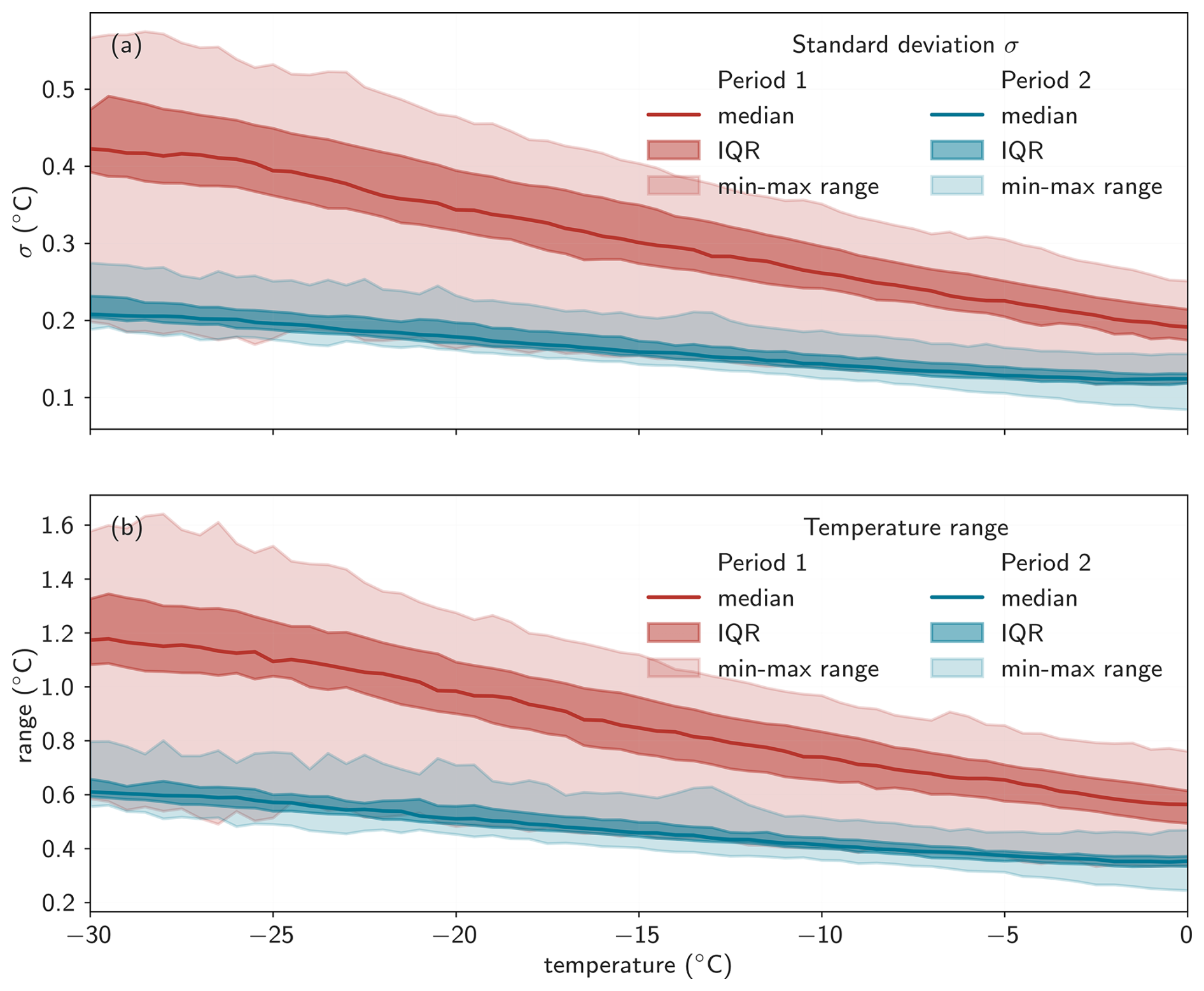
Figure A4Temperature standard deviation and total temperature spread as a function of temperature. Median, interquartile range (IQR) and minimum-maximum range temperature of (a) standard deviations and (b) total temperature range (Tmax–Tmin) derived from eight PT100 temperature sensors across 212 experiments, separately for period 1 and period 2. Both panels illustrate the variability in temperature homogeneity within the aluminum block as a function of temperature.
As these changes affect temperature stability, the sample measurement period is separated into period 1 and period 2 (shaded regions in Fig. A3), for which temperature uncertainty is quantified independently. Figure A4 shows the median, interquartile range, and full range of temperature standard deviations and total temperature spread across the eight sensors as a function of temperature for both periods. Temperature variability increases toward colder temperatures in both cases, but more strongly in period 1. At −30 °C, the maximum standard deviation reaches 0.57 °C in period 1, compared to 0.28 °C in period 2, which is similar to comparable set-ups (e.g., 0.6 °C in David et al., 2019; 0.5 °C in Miller et al., 2021). The median total temperature spread across the block reaches 1.2 and 0.6 °C for period 1 and period 2, respectively.
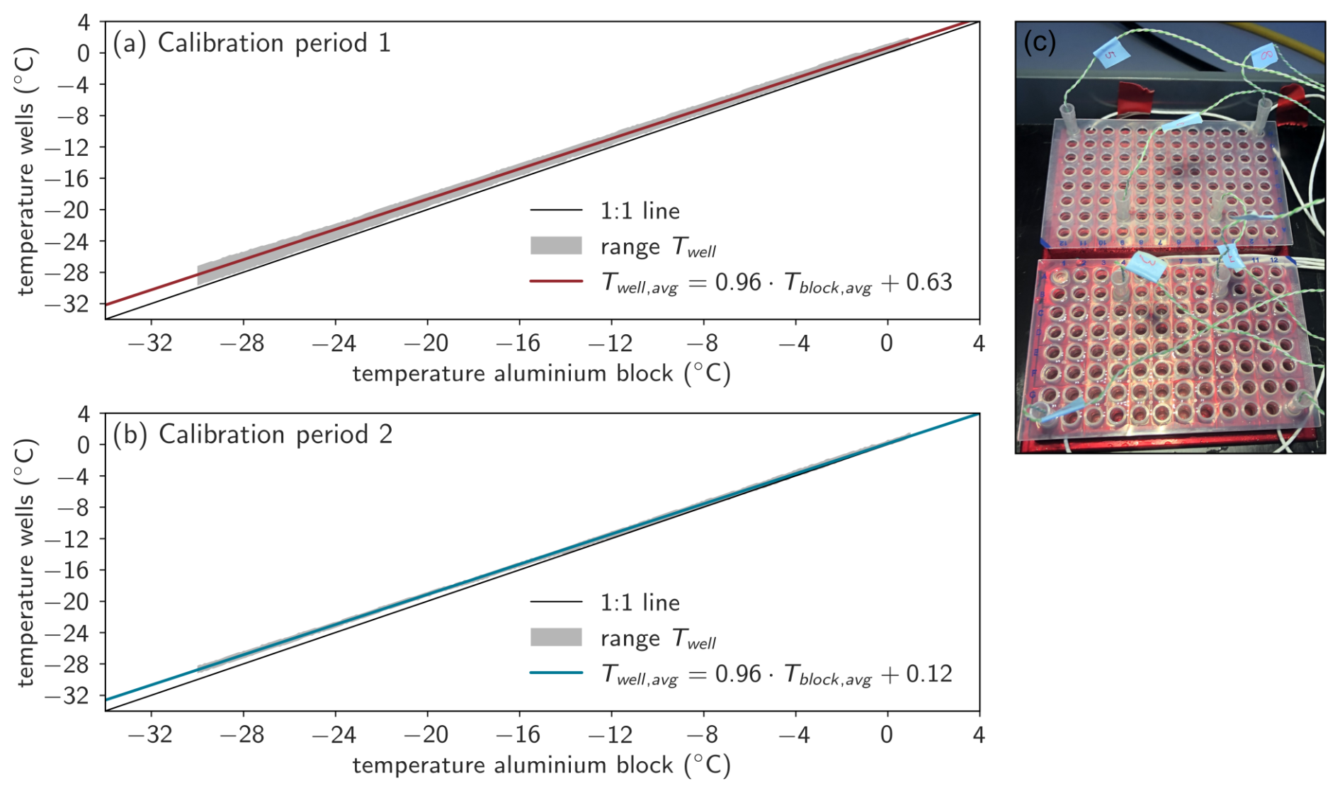
Figure A5SPICE temperature calibration. (a) Calibration for period 1, conducted with airflow and under warmer laboratory conditions, and (b) for period 2, conducted without airflow and under colder laboratory conditions. For each period, three calibration experiments with different thermocouple placements were performed. Grey shading indicates the range of temperatures measured in the wells, while the linear fit represents the mean slope and intercept derived from the three calibration experiments. (c) Experimental setup illustrating the thermocouples inserted into the wells of the PCR trays.
Some heat loss is expected between the temperature sensors embedded in the aluminum block and the actual droplet positions in the PCR trays. To quantify this offset, temperature calibration experiments were carried out using the following setup: eight K-type thermocouples (TW-KT3P-1000, Thermosense, UK) were placed into wells filled with ethanol (Fig. A5c), and a standard freezing experiment was performed with a cooling rate of 0.33 °C min−1. Thermocouple temperatures were recorded using an external data logger (HH-4208SD, Thermosense, UK). A linear regression was applied between the average temperature measured by the PT100 sensors in the block (Tblock,avg) and the average temperature in the wells measured by the thermocouples (Twell,avg).
Given the temperature variability over time, which also affects differences in block and well temperatures, we applied separate calibrations for period 1 and period 2. For period 1, three calibration experiments were conducted with filtered airflow over the glass pane, using different thermocouple placements to ensure representative spatial coverage across the PCR trays. The combined calibration (Fig. A5a, average of slopes and intercepts of the three experiments) yields the relationship:
The calibration uncertainty is approximated by the standard deviations of slopes (σm: ±0.008) and intercepts (σb: ±0.26). Combined with the maximum block-temperature uncertainty (: ±0.57), the uncertainty in well temperatures is estimated by propagating the uncertainties:
For period 1, this results in a total well-temperature uncertainty of °C.
For period 2, three additional calibration experiments were performed with the same setup, but without the filtered airflow. The combined calibration (Fig. A5b) results in the following equation:
The smaller offset between block and well temperatures (0.51 °C smaller than for period 1), together with reduced temperature variability, leads to a lower total uncertainty of °C, based on Eq. (A10) (σm: ±0.001, maximum : ±0.28, σb: ±0.054). Although Eq. (A10) assumes independent uncertainties, which may not strictly be true, the resulting estimates remain conservative because we use the maximum observed temperature standard deviations across all experiments.
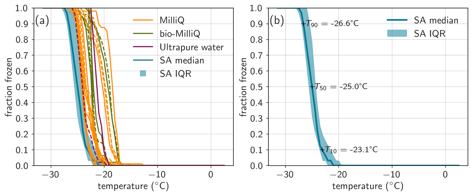
Figure A6Background freezing. (a) Frozen fraction of different purified water types: Milli-Q (Millipak®, Merck, Germany), bio-Milli-Q (Biopak®, Merck, Germany), and Ultrapure water (Cat. No. 022934.K2, Thermo Scientific, USA), shown either unfiltered (solid line) or filtered through a 0.1 µm syringe filter (Whatman® Puradisc 25 TF, dashed lines), and molecular-biology-free reagent water from Sigma Aldrich (SA) water (median and IQR of 152 experiments). (b) Median and IQR of SA water including freezing temperatures where a fraction of 0.1, 0.5, and 0.9 of droplets are frozen (T10, T50, T90).
A2.2 Characteristic background freezing temperature
All experiments include negative controls of the background water to quantify potential background freezing events caused by impurities in the water, contamination during handling, or the PCR tray material itself. We use SA water for our experiments, which has previously been shown to exhibit low and more reproducible freezing behavior than other purified waters (David et al., 2019; Miller et al., 2021), consistent with our comparison in the laboratory (Fig. A6a). We define the characteristic background freezing temperature of the set-up as the median of the median freezing temperature (T50, where frozen fraction = 0.5) of 152 experiments (Fig. A6b, including both dedicated water testing experiments and negative controls within sample freezing assays), which is −24.71 °C (interquartile range of 0.88 °C) for 50 µL droplets. This characteristic background freezing temperature is therefore similar to other INP freezing set-ups (e.g., Chen et al., 2018; David et al., 2019; Miller et al., 2021). For the negative controls within sample freezing assays, we typically use the same PCR tray region, which might introduce a small bias, though it is below the reported temperature uncertainty. The cumulative INP concentrations reported for samples are corrected with the water background of each individual experiment, as described in Eq. (A4).
A2.3 Validation experiments
To validate SPICE, we tested two commercially available materials with published ice-nucleation freezing spectra, NX-illite and lignin. NX-illite is a clay mineral with well-documented freezing activity (e.g., Beall et al., 2017; David et al., 2019; Harrison et al., 2018; Hiranuma et al., 2015; Miller et al., 2021) and can be representative of atmospheric dust which contains a high illite fraction (approx. 40 %, Broadley et al., 2012; Murray et al., 2012). Following David et al. (2019) and Miller et al. (2021), suspensions of 0.1, 0.05, and 0.001 g L−1 NX-illite (Adolf Gottfried Tonwerke GmbH, Germany) were prepared using SA water, and ns,BET was calculated with Eq. (A7) using a Brunauer-Emmett-Teller (BET) specific surface area of 124.4 m2 g−1 for NX-illite (Hiranuma et al., 2015). The resulting ns,BET spectra are shown in Fig. A7, including results obtained with the same concentrations from DRINCZ, FINC, and LINDA (Miller et al., 2021), as well as measurements from IR-NIPI (Harrison et al., 2018), SIO-AIS (Beall et al., 2017), and the range and wet suspension parameterization from the intercomparison campaign described in Hiranuma et al. (2015). The parameterization by Hiranuma et al. (2015) agrees reasonably well with the SPICE measurements around −15 °C, but predicts ns,BET values 2–10 times higher at colder temperatures. The SPICE ns,BET values fall within the broad range reported by Hiranuma et al. (2015), though generally towards the lower end. They are also 1–2 orders of magnitude lower than NX-illite ns,BET values measured by Beall et al. (2017) and Harrison et al. (2018).
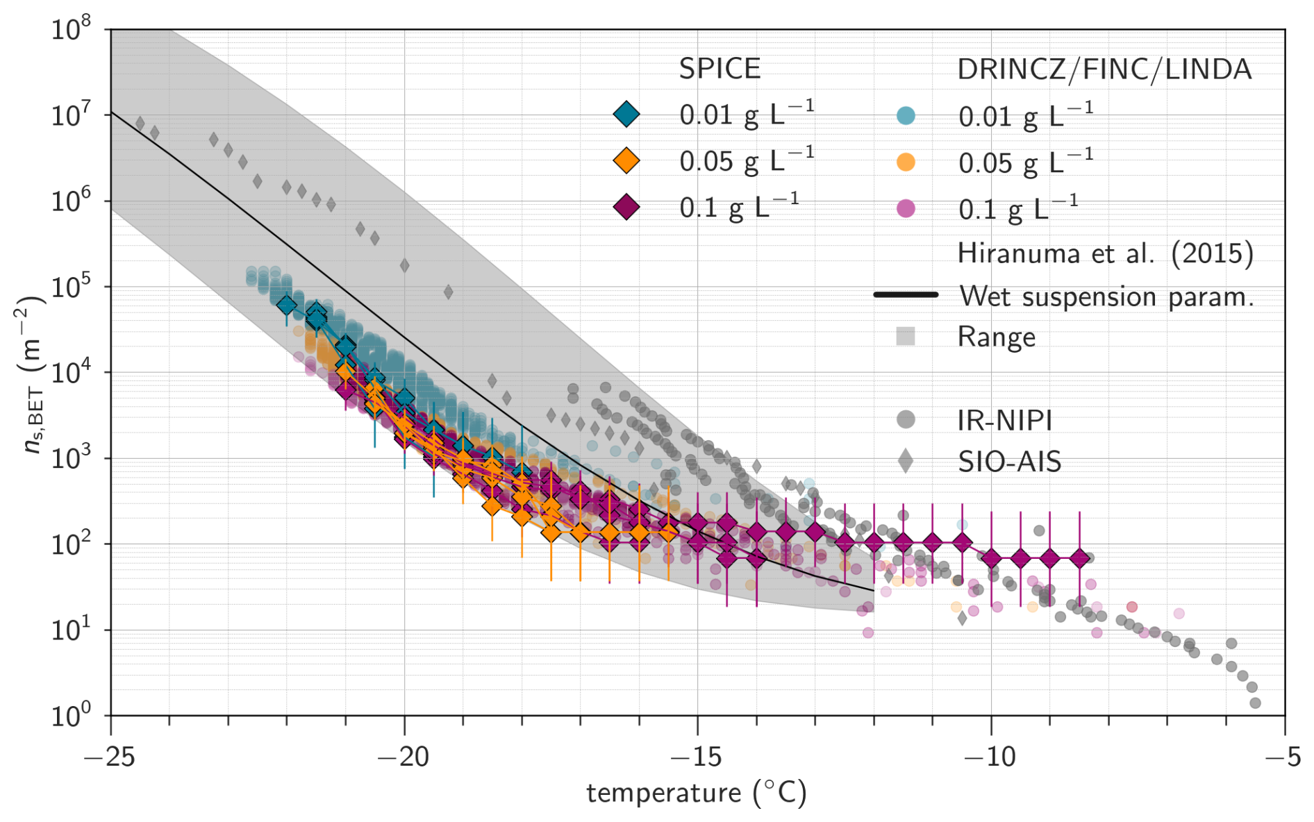
Figure A7NX-illite comparison. Ice-active site densities per surface area (ns,BET) of NX-illite suspensions with concentrations of 0.01, 0.05, and 0.1 g L−1 measured with SPICE. ns,BET was calculated using a BET surface area of 124.4 m2 g−1 for NX-illite (Hiranuma et al., 2015). For comparison, results obtained with the same concentrations from DRINCZ, FINC, and LINDA (Miller et al., 2021), as well as measurements from IR-NIPI (Harrison et al., 2018) and SIO-AIS (Beall et al., 2017), are shown. The shaded region and solid line indicate the range and wet-suspension parameterization from Hiranuma et al. (2015).
SPICE measurements are in better agreement with ns,BET spectra reported in Miller et al. (2021), but depend on both temperature and particle concentration. For suspensions of 0.01 and 0.05 g L−1, the SPICE measurements agree well with Miller et al. (2021) at temperatures colder than about −21 and −18 °C, respectively, but are around a factor of two lower at higher temperatures. At 0.1 g L−1, the measurements match closely below −14 °C, but SPICE shows up to two times higher ns,BET at warmer temperatures.
Despite the large variability in published NX-illite freezing spectra, the SPICE measurements lie within the overall reported range and show broadly consistent behavior. The spread among different datasets could reflect variability introduced by differences in sample properties, e.g., including particle dispersion, sedimentation, and size distribution characteristics, as well as differences in sample preparation and measurement methods (Hiranuma et al., 2015; Miller et al., 2021). Unlike the coordinated comparisons in Miller et al. (2021) and Hiranuma et al. (2015), the NX-illite sample used in this study was purchased independently, which may further contribute to deviations between datasets.
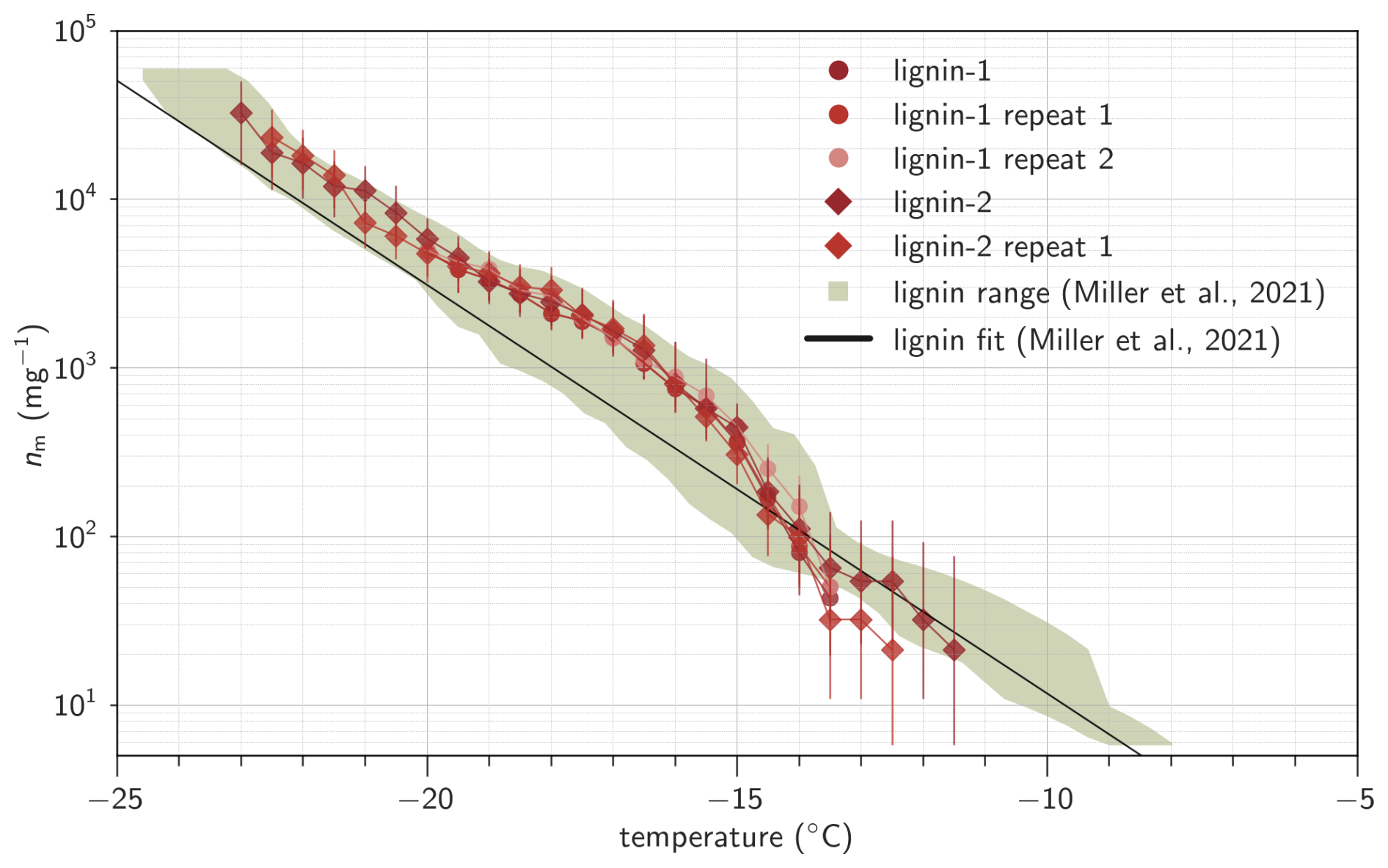
Figure A8Lignin comparison. Ice-active site densities per mass (nm) of lignin suspensions measured with SPICE. The first suspension (lignin-1) with 20 mg C L−1 solution was measured three times. The second suspension (lignin-2) was measured twice, including the solution of 20 mg C L−1 and a 10-fold dilution. The shaded region and solid line indicate the range and parameterization from Miller et al. (2021).
Commercial lignin has been proposed by Miller et al. (2021) as an ice-nucleation reference material because it showed better agreement among three different droplet-freezing instruments compared to NX-illite, displayed consistent results across production batches, and remained stable over time. Lignin is a natural, biosphere-abundant polymer, and the commercial material used here is sourced as a by-product of the pulp and paper industry. For this study, we used alkali low-sulfonate kraft lignin (CAS 8068-05-1, Sigma Aldrich, product code 471003) to prepare suspensions equivalent to 20 g C L−1, and calculated the ice-nucleating active site density per unit carbon mass (nm) following Miller et al. (2021):
where Cm(TOC) is the mass concentration of carbon in the solution. For the lignin used here, this concentration is determined from the lignin mass (40 mg L−1) and the manufacturer-reported carbon content (50.3 %).
The SPICE freezing spectra of lignin show high reproducibility across repeated experiments, and are in good agreement with the results reported by Miller et al. (2021). While the parameterization based on multiple instruments shows up to a factor of 2–3 difference in the −15 to −20 °C temperature range, the SPICE measurements closely match the batch-specific measurements in Miller et al. (2021, upper green range in Fig. A8). In addition, we participated in an offline INP comparison initiative coordinated by the Centre for Cloud Ice nucleation (CCIce) within the Aerosol, Clouds, Trace Gases Research Infrastructure (ACTRIS) framework in 2025. SPICE compared well to the other instruments and fell within the statistical uncertainty of the ensemble of measurements (Lacher and Höhler, 2025).
Overall, the high reproducibility of our measurements and their good agreement with published data for NX-illite, lignin, and other droplet freezing instruments demonstrate that SPICE provides robust and reliable measurements of immersion-mode INPs.
INP data are available at https://doi.org/10.5281/zenodo.18375165 (Bergner et al., 2026a) and https://doi.org/10.5281/zenodo.18376580 (Bergner et al., 2026b). Meteorological data are available at https://doi.org/10.5281/zenodo.15388080 (Alden et al., 2025). Aerosol data are available at https://doi.org/10.5281/zenodo.15387340 (Bergner et al., 2025a) and https://doi.org/10.5281/zenodo.15388013 (Bergner et al., 2025b).
The supplement related to this article is available online at https://doi.org/10.5194/acp-26-10557-2026-supplement.
NB, GM, JA, IA, LB, DF, LF, BH, JW and JS took part in the field campaign to collect samples and perform measurements. JS conceptualized the campaign and acquired funding. AB, KH, LL and KB provided their expertise on INP measurements. RP and LF built the INP filter sampler. NB analyzed the samples with help from CG. GM conducted microbiology measurements. CA and NB collected and analyzed dust samples in the Alps. NB analyzed the data and wrote the manuscript with contributions and feedback from all authors.
The contact author has declared that none of the authors has any competing interests.
Publisher's note: Copernicus Publications remains neutral with regard to jurisdictional claims made in the text, published maps, institutional affiliations, or any other geographical representation in this paper. The authors bear the ultimate responsibility for providing appropriate place names. Views expressed in the text are those of the authors and do not necessarily reflect the views of the publisher.
J.S. holds the Ingvar Kamprad chair for Extreme Environments Research sponsored by Ferring Pharmaceuticals. We thank the Narsaq International Research Station team, especially Lise Autogena, and the residents of Narsaq and Narsarsuaq for their generosity, curiosity, and close collaboration throughout our work. We are grateful to the Kujalleq commune for their support and for granting the land-use permissions that made this research possible. We recognize that our study took place on the traditional lands of the people of Kalaallit Nunaat and deeply appreciate the opportunity to carry out this research in a spirit of respect and cooperation with the community. We would like to thank Son Pham-Ba and Evan Thomas for the development of the SPICE image analysis and database. We thank Pascal Schouwink for the XRD measurements, and Emad Oveisi for SEM-EDX measurements.
This research has been supported by the Schweizerischer Nationalfonds zur Förderung der Wissenschaftlichen Forschung (grant no. 200021_212101), the Swiss Polar Institute (grant no. SPI-FLAG-2021-002), the École Polytechnique Fédérale de Lausanne (grant no. ENAC Flagship 2022 ECO-Plains), and the HORIZON EUROPE European Research Council (grant no. 101003826).
This paper was edited by Markus Petters and reviewed by three anonymous referees.
Agresti, A. and Coull, B. A.: Approximate Is Better than “Exact” for Interval Estimation of Binomial Proportions, Am. Stat., 52, 119–126, https://doi.org/10.2307/2685469, 1998.
Alden, J., Bergner, N., Heutte, B., Farinotti, D., Favre, L., and Schmale, J.: Meteorological data from multiple weather stations in Narsarsuaq and Narsaq, South Greenland during Greenfjord 2023, Zenodo [data set], https://doi.org/10.5281/zenodo.15388080, 2025.
Alden, J., Bergner, N., Heutte, B., Favre, L., Surdu, M., Weng, J., Augugliaro, M., Winiger, P., Dönmez, B., Pohorsky, R., Calmer, R., Chatterjee, C., Violaki, K., Nenes, A., Gregor, L., Henning, S., and Schmale, J.: Vertically-resolved source contributions to climate-relevant aerosol properties in Southern Greenlandic fjord systems, Atmos. Chem. Phys., 26, 7165–7191, https://doi.org/10.5194/acp-26-7165-2026, 2026.
Atkinson, J. D., Murray, B. J., Woodhouse, M. T., Whale, T. F., Baustian, K. J., Carslaw, K. S., Dobbie, S., O'Sullivan, D., and Malkin, T. L.: The importance of feldspar for ice nucleation by mineral dust in mixed-phase clouds, Nature, 498, 355–358, https://doi.org/10.1038/nature12278, 2013.
Barr, S. L., Wyld, B., McQuaid, J. B., Neely Iii, R. R., and Murray, B. J.: Southern Alaska as a source of atmospheric mineral dust and ice-nucleating particles, Sci. Adv., 9, eadg3708, https://doi.org/10.1126/sciadv.adg3708, 2023.
Barry, K. R., Hill, T. C. J., Jentzsch, C., Moffett, B. F., Stratmann, F., and DeMott, P. J.: Pragmatic protocols for working cleanly when measuring ice nucleating particles, Atmos. Res., 250, 105419, https://doi.org/10.1016/j.atmosres.2020.105419, 2021.
Barry, K. R., Hill, T. C. J., Nieto-Caballero, M., Douglas, T. A., Kreidenweis, S. M., DeMott, P. J., and Creamean, J. M.: Active thermokarst regions contain rich sources of ice-nucleating particles, Atmos. Chem. Phys., 23, 15783–15793, https://doi.org/10.5194/acp-23-15783-2023, 2023.
Barry, K. R., Hill, T. C. J., Kreidenweis, S. M., DeMott, P. J., Tobo, Y., and Creamean, J. M.: Bioaerosols as indicators of central Arctic ice nucleating particle sources, Atmos. Chem. Phys., 25, 11919–11933, https://doi.org/10.5194/acp-25-11919-2025, 2025.
Beall, C. M., Stokes, M. D., Hill, T. C., DeMott, P. J., DeWald, J. T., and Prather, K. A.: Automation and heat transfer characterization of immersion mode spectroscopy for analysis of ice nucleating particles, Atmos. Meas. Tech., 10, 2613–2626, https://doi.org/10.5194/amt-10-2613-2017, 2017.
Beall, C. M., Lucero, D., Hill, T. C., DeMott, P. J., Stokes, M. D., and Prather, K. A.: Best practices for precipitation sample storage for offline studies of ice nucleation in marine and coastal environments, Atmos. Meas. Tech., 13, 6473–6486, https://doi.org/10.5194/amt-13-6473-2020, 2020.
Beck, I., Moallemi, A., Heutte, B., Pernov, J. B., Bergner, N., Rolo, M., Quéléver, L. L. J., Laurila, T., Boyer, M., Jokinen, T., Angot, H., Hoppe, C. J. M., Müller, O., Creamean, J., Frey, M. M., Freitas, G., Zinke, J., Salter, M., Zieger, P., Mirrielees, J. A., Kempf, H. E., Ault, A. P., Pratt, K. A., Gysel-Beer, M., Henning, S., Tatzelt, C., and Schmale, J.: Characteristics and sources of fluorescent aerosols in the central Arctic Ocean, Elem. Sci. Anth., 12, 00125, https://doi.org/10.1525/elementa.2023.00125, 2024.
Bergner, N., Alden, J., Heutte, B., Weng, J., Violaki, K., Favre, L., and Schmale, J.: Fidas measurements of total aerosol number concentration and number size distributions in summertime in Narsaq, South Greenland during Greenfjord 2023, Zenodo [data set], https://doi.org/10.5281/zenodo.15387340, 2025a.
Bergner, N., Alden, J., Heutte, B., Favre, L., Farinotti, D., and Schmale, J.: Portable Optical Particle Sizer (POPS) measurements of aerosol particle number concentration and size distribution in summertime in Narsarsuaq and Narsaq, South Greenland during Greenfjord 2023, Zenodo [data set], https://doi.org/10.5281/zenodo.15388013, 2025b.
Bergner, N., Marsh, G., Altshuler, I., Bröder, L., Farinotti, D., Guillosson, C., and Schmale, J.: Ice-nucleating activity of glacially sourced dust collected in glacial outwash plains in Narsarsuaq, Igaliku, and Kangerlussuaq, Greenland, Zenodo [data set], https://doi.org/10.5281/zenodo.18375165, 2026a.
Bergner, N., Alden, J., Farinotti, D., Favre, L., Heutte, B., Pohorsky, R., Weng, J., and Schmale, J.: Ice-nucleating particle concentrations from June–August 2023 in Narsaq and Narsarsuaq in southern Greenland, Zenodo [data set], https://doi.org/10.5281/zenodo.18376580, 2026b.
Böhmländer, A., Lacher, L., Höhler, K., Brus, D., Doulgeris, K.-M., Girdwood, J., Leisner, T., and Möhler, O.: Measurement of the ice-nucleating particle concentration using a mobile filter-based sampler on-board of a fixed-wing uncrewed aerial vehicle during the Pallas Cloud Experiment 2022, Earth Syst. Sci. Data, 17, 6157–6164, https://doi.org/10.5194/essd-17-6157-2025, 2025.
Boose, Y., Welti, A., Atkinson, J., Ramelli, F., Danielczok, A., Bingemer, H. G., Plötze, M., Sierau, B., Kanji, Z. A., and Lohmann, U.: Heterogeneous ice nucleation on dust particles sourced from nine deserts worldwide – Part 1: Immersion freezing, Atmos. Chem. Phys., 16, 15075–15095, https://doi.org/10.5194/acp-16-15075-2016, 2016.
Bradley, J. A., Arndt, S., Šabacká, M., Benning, L. G., Barker, G. L., Blacker, J. J., Yallop, M. L., Wright, K. E., Bellas, C. M., Telling, J., Tranter, M., and Anesio, A. M.: Microbial dynamics in a High Arctic glacier forefield: a combined field, laboratory, and modelling approach, Biogeosciences, 13, 5677–5696, https://doi.org/10.5194/bg-13-5677-2016, 2016.
Broadley, S. L., Murray, B. J., Herbert, R. J., Atkinson, J. D., Dobbie, S., Malkin, T. L., Condliffe, E., and Neve, L.: Immersion mode heterogeneous ice nucleation by an illite rich powder representative of atmospheric mineral dust, Atmos. Chem. Phys., 12, 287–307, https://doi.org/10.5194/acp-12-287-2012, 2012.
Brown, L. D., Cai, T. T., and DasGupta, A.: Interval Estimation for a Binomial Proportion, Statist. Sci., 16, https://doi.org/10.1214/ss/1009213286, 2001.
Bullard, J. E. and Mockford, T.: Seasonal and decadal variability of dust observations in the Kangerlussuaq area, west Greenland, Arct. Antarct. Alp. Res., 50, S100011, https://doi.org/10.1080/15230430.2017.1415854, 2018.
Bullard, J. E., Baddock, M., Bradwell, T., Crusius, J., Darlington, E., Gaiero, D., Gassó, S., Gisladottir, G., Hodgkins, R., McCulloch, R., McKenna-Neuman, C., Mockford, T., Stewart, H., and Thorsteinsson, T.: High-latitude dust in the Earth system, Rev. Geophys., 54, 447–485, https://doi.org/10.1002/2016RG000518, 2016.
Bullard, J. E., Prater, C., Baddock, M. C., and Anderson, N. J.: Diurnal and seasonal source-proximal dust concentrations in complex terrain, West Greenland, Earth Surf. Proc. Land., 48, 2808–2827, https://doi.org/10.1002/esp.5661, 2023.
Carlson, D. F., Pavalko, W. J., Petersen, D., Olsen, M., and Hass, A. E.: Maker Buoy Variants for Water Level Monitoring and Tracking Drifting Objects in Remote Areas of Greenland, Sensors, 20, 1254, https://doi.org/10.3390/s20051254, 2020.
Carrivick, J. L. and Tweed, F. S.: A review of glacier outburst floods in Iceland and Greenland with a megafloods perspective, Earth-Sci. Rev., 196, 102876, https://doi.org/10.1016/j.earscirev.2019.102876, 2019.
Ceppi, P., Brient, F., Zelinka, M. D., and Hartmann, D. L.: Cloud feedback mechanisms and their representation in global climate models, WIREs Climate Change, 8, e465, https://doi.org/10.1002/wcc.465, 2017.
Chen, J., Wu, Z., Augustin-Bauditz, S., Grawe, S., Hartmann, M., Pei, X., Liu, Z., Ji, D., and Wex, H.: Ice-nucleating particle concentrations unaffected by urban air pollution in Beijing, China, Atmos. Chem. Phys., 18, 3523–3539, https://doi.org/10.5194/acp-18-3523-2018, 2018.
Chen, J., Wu, Z., Gong, X., Qiu, Y., Chen, S., Zeng, L., and Hu, M.: Anthropogenic Dust as a Significant Source of Ice-Nucleating Particles in the Urban Environment, Earths Future, 12, e2023EF003738, https://doi.org/10.1029/2023EF003738, 2024.
Conen, F. and Einbock, A.: Release of ice-nucleating particles from leaves during rainfall, Sci. Nat., 112, 29, https://doi.org/10.1007/s00114-025-01980-6, 2025.
Conen, F., Morris, C. E., Leifeld, J., Yakutin, M. V., and Alewell, C.: Biological residues define the ice nucleation properties of soil dust, Atmos. Chem. Phys., 11, 9643–9648, https://doi.org/10.5194/acp-11-9643-2011, 2011.
Crusius, J., Schroth, A. W., Gasso, S., Moy, C. M., Levy, R. C., and Gatica, M.: Glacial flour dust storms in the Gulf of Alaska: hydrologic and meteorological controls and their importance as a source of bioavailable iron, Geophys. Res. Lett., 38, https://doi.org/10.1029/2010GL046573, 2011.
Dagsson-Waldhauserova, P., Arnalds, O., and Olafsson, H.: Long-term frequency and characteristics of dust storm events in Northeast Iceland (1949–2011), Atmos. Environ., 77, 117–127, https://doi.org/10.1016/j.atmosenv.2013.04.075, 2013.
Daily, M. I., Tarn, M. D., Whale, T. F., and Murray, B. J.: An evaluation of the heat test for the ice-nucleating ability of minerals and biological material, Atmos. Meas. Tech., 15, 2635–2665, https://doi.org/10.5194/amt-15-2635-2022, 2022.
David, R. O., Cascajo-Castresana, M., Brennan, K. P., Rösch, M., Els, N., Werz, J., Weichlinger, V., Boynton, L. S., Bogler, S., Borduas-Dedekind, N., Marcolli, C., and Kanji, Z. A.: Development of the DRoplet Ice Nuclei Counter Zurich (DRINCZ): validation and application to field-collected snow samples, Atmos. Meas. Tech., 12, 6865–6888, https://doi.org/10.5194/amt-12-6865-2019, 2019.
DeMott, P. J., Cziczo, D. J., Prenni, A. J., Murphy, D. M., Kreidenweis, S. M., Thomson, D. S., Borys, R., and Rogers, D. C.: Measurements of the concentration and composition of nuclei for cirrus formation, P. Natl. Acad. Sci. USA, 100, 14655–14660, https://doi.org/10.1073/pnas.2532677100, 2003.
DeMott, P. J., Prenni, A. J., Liu, X., Kreidenweis, S. M., Petters, M. D., Twohy, C. H., Richardson, M. S., Eidhammer, T., and Rogers, D. C.: Predicting global atmospheric ice nuclei distributions and their impacts on climate, P. Natl. Acad. Sci. USA, 107, 11217–11222, https://doi.org/10.1073/pnas.0910818107, 2010.
DeMott, P. J., Prenni, A. J., McMeeking, G. R., Sullivan, R. C., Petters, M. D., Tobo, Y., Niemand, M., Möhler, O., Snider, J. R., Wang, Z., and Kreidenweis, S. M.: Integrating laboratory and field data to quantify the immersion freezing ice nucleation activity of mineral dust particles, Atmos. Chem. Phys., 15, 393–409, https://doi.org/10.5194/acp-15-393-2015, 2015.
DeMott, P. J., Hill, T. C. J., McCluskey, C. S., Prather, K. A., Collins, D. B., Sullivan, R. C., Ruppel, M. J., Mason, R. H., Irish, V. E., Lee, T., Hwang, C. Y., Rhee, T. S., Snider, J. R., McMeeking, G. R., Dhaniyala, S., Lewis, E. R., Wentzell, J. J. B., Abbatt, J., Lee, C., Sultana, C. M., Ault, A. P., Axson, J. L., Diaz Martinez, M., Venero, I., Santos-Figueroa, G., Stokes, M. D., Deane, G. B., Mayol-Bracero, O. L., Grassian, V. H., Bertram, T. H., Bertram, A. K., Moffett, B. F., and Franc, G. D.: Sea spray aerosol as a unique source of ice nucleating particles, P. Natl. Acad. Sci. USA, 113, 5797–5803, https://doi.org/10.1073/pnas.1514034112, 2016.
DeMott, P. J., Hill, T. C. J., Petters, M. D., Bertram, A. K., Tobo, Y., Mason, R. H., Suski, K. J., McCluskey, C. S., Levin, E. J. T., Schill, G. P., Boose, Y., Rauker, A. M., Miller, A. J., Zaragoza, J., Rocci, K., Rothfuss, N. E., Taylor, H. P., Hader, J. D., Chou, C., Huffman, J. A., Pöschl, U., Prenni, A. J., and Kreidenweis, S. M.: Comparative measurements of ambient atmospheric concentrations of ice nucleating particles using multiple immersion freezing methods and a continuous flow diffusion chamber, Atmos. Chem. Phys., 17, 11227–11245, https://doi.org/10.5194/acp-17-11227-2017, 2017.
Diehl, K., Matthias-Maser, S., Jaenicke, R., and Mitra, S. K.: The ice nucleating ability of pollen:: Part II. Laboratory studies in immersion and contact freezing modes, Atmos. Res., 61, 125–133, https://doi.org/10.1016/S0169-8095(01)00132-6, 2002.
Donhauser, J. and Frey, B.: Alpine soil microbial ecology in a changing world, FEMS Microbiol. Ecol., 94, https://doi.org/10.1093/femsec/fiy099, 2018.
Dreischmeier, K., Budke, C., Wiehemeier, L., Kottke, T., and Koop, T.: Boreal pollen contain ice-nucleating as well as ice-binding “antifreeze” polysaccharides, Sci. Rep., 7, 41890, https://doi.org/10.1038/srep41890, 2017.
Duan, P., Hu, W., Wu, Z., Bi, K., Zhu, J., and Fu, P.: Ice nucleation activity of airborne pollen: A short review of results from laboratory experiments, Atmos. Res., 285, 106659, https://doi.org/10.1016/j.atmosres.2023.106659, 2023.
Failor, K. C., Schmale III, D. G., Vinatzer, B. A., and Monteil, C. L.: Ice nucleation active bacteria in precipitation are genetically diverse and nucleate ice by employing different mechanisms, ISME J., 11, 2740–2753, https://doi.org/10.1038/ismej.2017.124, 2017.
Field, P. R., Lawson, R. P., Brown, P. R. A., Lloyd, G., Westbrook, C., Moisseev, D., Miltenberger, A., Nenes, A., Blyth, A., Choularton, T., Connolly, P., Buehl, J., Crosier, J., Cui, Z., Dearden, C., DeMott, P., Flossmann, A., Heymsfield, A., Huang, Y., Kalesse, H., Kanji, Z. A., Korolev, A., Kirchgaessner, A., Lasher-Trapp, S., Leisner, T., McFarquhar, G., Phillips, V., Stith, J., and Sullivan, S.: Secondary Ice Production: Current State of the Science and Recommendations for the Future, Meteor. Mon., 58, 7.1–7.20, https://doi.org/10.1175/AMSMONOGRAPHS-D-16-0014.1, 2017.
Fröhlich-Nowoisky, J., Hill, T. C. J., Pummer, B. G., Yordanova, P., Franc, G. D., and Pöschl, U.: Ice nucleation activity in the widespread soil fungus Mortierella alpina, Biogeosciences, 12, 1057–1071, https://doi.org/10.5194/bg-12-1057-2015, 2015.
Fröhlich-Nowoisky, J., Kampf, C. J., Weber, B., Huffman, J. A., Pöhlker, C., Andreae, M. O., Lang-Yona, N., Burrows, S. M., Gunthe, S. S., Elbert, W., Su, H., Hoor, P., Thines, E., Hoffmann, T., Després, V. R., and Pöschl, U.: Bioaerosols in the Earth system: Climate, health, and ecosystem interactions, Atmos. Res., 182, 346–376, https://doi.org/10.1016/j.atmosres.2016.07.018, 2016.
Frossard, A., Hammes, F., and Gessner, M. O.: Flow Cytometric Assessment of Bacterial Abundance in Soils, Sediments and Sludge, Front. Microbiol., 7, https://doi.org/10.3389/fmicb.2016.00903, 2016.
Górniak, D., Marszałek, H., Kwaśniak-Kominek, M., Rzepa, G., and Manecki, M.: Soil formation and initial microbiological activity on a foreland of an Arctic glacier (SW Svalbard), Appl. Soil Ecol., 114, 34–44, https://doi.org/10.1016/j.apsoil.2017.02.017, 2017.
Gratzl, J., Böhmländer, A., Pätsi, S., Pogner, C.-E., Gorfer, M., Brus, D., Doulgeris, K. M., Wieland, F., Asmi, E., Saarto, A., Möhler, O., Stolzenburg, D., and Grothe, H.: Locally emitted fungal spores serve as high-temperature ice nucleating particles in the European sub-Arctic, Atmos. Chem. Phys., 25, 12007–12035, https://doi.org/10.5194/acp-25-12007-2025, 2025.
Grimes, M., Carrivick, J. L., Smith, M. W., and Comber, A. J.: Land cover changes across Greenland dominated by a doubling of vegetation in three decades, Sci. Rep., 14, 3120, https://doi.org/10.1038/s41598-024-52124-1, 2024.
Groot Zwaaftink, C. D., Grythe, H., Skov, H., and Stohl, A.: Substantial contribution of northern high-latitude sources to mineral dust in the Arctic, J. Geophys. Res.-Atmos., 121, 13678–13697, https://doi.org/10.1002/2016JD025482, 2016.
Hamzehpour, N., Marcolli, C., Pashai, S., Klumpp, K., and Peter, T.: Measurement report: The Urmia playa as a source of airborne dust and ice-nucleating particles – Part 1: Correlation between soils and airborne samples, Atmos. Chem. Phys., 22, 14905–14930, https://doi.org/10.5194/acp-22-14905-2022, 2022.
Hanna, J. W., Schultz, D. M., and Irving, A. R.: Cloud-Top Temperatures for Precipitating Winter Clouds, J. Appl. Meteorol. Clim., 47, 351–359, https://doi.org/10.1175/2007JAMC1549.1, 2008.
Hara, K., Maki, T., Kakikawa, M., Kobayashi, F., and Matsuki, A.: Effects of different temperature treatments on biological ice nuclei in snow samples, Atmos. Environ., 140, 415–419, https://doi.org/10.1016/j.atmosenv.2016.06.011, 2016.
Harrison, A. D., Whale, T. F., Rutledge, R., Lamb, S., Tarn, M. D., Porter, G. C. E., Adams, M. P., McQuaid, J. B., Morris, G. J., and Murray, B. J.: An instrument for quantifying heterogeneous ice nucleation in multiwell plates using infrared emissions to detect freezing, Atmos. Meas. Tech., 11, 5629–5641, https://doi.org/10.5194/amt-11-5629-2018, 2018.
Harrison, A. D., Lever, K., Sanchez-Marroquin, A., Holden, M. A., Whale, T. F., Tarn, M. D., McQuaid, J. B., and Murray, B. J.: The ice-nucleating ability of quartz immersed in water and its atmospheric importance compared to K-feldspar, Atmos. Chem. Phys., 19, 11343–11361, https://doi.org/10.5194/acp-19-11343-2019, 2019.
Hill, T. C. J., DeMott, P. J., Tobo, Y., Fröhlich-Nowoisky, J., Moffett, B. F., Franc, G. D., and Kreidenweis, S. M.: Sources of organic ice nucleating particles in soils, Atmos. Chem. Phys., 16, 7195–7211, https://doi.org/10.5194/acp-16-7195-2016, 2016.
Hiranuma, N., Augustin-Bauditz, S., Bingemer, H., Budke, C., Curtius, J., Danielczok, A., Diehl, K., Dreischmeier, K., Ebert, M., Frank, F., Hoffmann, N., Kandler, K., Kiselev, A., Koop, T., Leisner, T., Möhler, O., Nillius, B., Peckhaus, A., Rose, D., Weinbruch, S., Wex, H., Boose, Y., DeMott, P. J., Hader, J. D., Hill, T. C. J., Kanji, Z. A., Kulkarni, G., Levin, E. J. T., McCluskey, C. S., Murakami, M., Murray, B. J., Niedermeier, D., Petters, M. D., O'Sullivan, D., Saito, A., Schill, G. P., Tajiri, T., Tolbert, M. A., Welti, A., Whale, T. F., Wright, T. P., and Yamashita, K.: A comprehensive laboratory study on the immersion freezing behavior of illite NX particles: a comparison of 17 ice nucleation measurement techniques, Atmos. Chem. Phys., 15, 2489–2518, https://doi.org/10.5194/acp-15-2489-2015, 2015.
Hoose, C. and Möhler, O.: Heterogeneous ice nucleation on atmospheric aerosols: a review of results from laboratory experiments, Atmos. Chem. Phys., 12, 9817–9854, https://doi.org/10.5194/acp-12-9817-2012, 2012.
Huang, Z., Huang, J., Hayasaka, T., Wang, S., Zhou, T., and Jin, H.: Short-cut transport path for Asian dust directly to the Arctic: a case study, Environ. Res. Lett., 10, 114018, https://doi.org/10.1088/1748-9326/10/11/114018, 2015.
Huffman, J. A., Prenni, A. J., DeMott, P. J., Pöhlker, C., Mason, R. H., Robinson, N. H., Fröhlich-Nowoisky, J., Tobo, Y., Després, V. R., Garcia, E., Gochis, D. J., Harris, E., Müller-Germann, I., Ruzene, C., Schmer, B., Sinha, B., Day, D. A., Andreae, M. O., Jimenez, J. L., Gallagher, M., Kreidenweis, S. M., Bertram, A. K., and Pöschl, U.: High concentrations of biological aerosol particles and ice nuclei during and after rain, Atmos. Chem. Phys., 13, 6151–6164, https://doi.org/10.5194/acp-13-6151-2013, 2013.
Irish, V. E., Hanna, S. J., Willis, M. D., China, S., Thomas, J. L., Wentzell, J. J. B., Cirisan, A., Si, M., Leaitch, W. R., Murphy, J. G., Abbatt, J. P. D., Laskin, A., Girard, E., and Bertram, A. K.: Ice nucleating particles in the marine boundary layer in the Canadian Arctic during summer 2014, Atmos. Chem. Phys., 19, 1027–1039, https://doi.org/10.5194/acp-19-1027-2019, 2019.
Jensen, L. Z., Simonsen, J. K., Pastor, A., Pearce, C., Nørnberg, P., Lund-Hansen, L. C., Finster, K., and Šantl-Temkiv, T.: Linking biogenic high-temperature ice nucleating particles in Arctic soils and streams to their microbial producers, Aerosol Research, 3, 81–100, https://doi.org/10.5194/ar-3-81-2025, 2025.
Joly, M., Attard, E., Sancelme, M., Deguillaume, L., Guilbaud, C., Morris, C. E., Amato, P., and Delort, A.-M.: Ice nucleation activity of bacteria isolated from cloud water, Atmos. Environ., 70, 392–400, https://doi.org/10.1016/j.atmosenv.2013.01.027, 2013.
Kanji, Z. A., Ladino, L. A., Wex, H., Boose, Y., Burkert-Kohn, M., Cziczo, D. J., and Krämer, M.: Overview of Ice Nucleating Particles, Meteor. Mon., 58, 1.1–1.33, https://doi.org/10.1175/AMSMONOGRAPHS-D-16-0006.1, 2017.
Kawai, K. and Matsui, H.: Contributions of Dust Source Regions to Ice Nucleating Particles in Mixed-Phase Clouds Simulated with a Global Climate–Aerosol Model, J. Climate, 38, 4925–4939, https://doi.org/10.1175/JCLI-D-24-0696.1, 2025.
Kawai, K., Matsui, H., and Tobo, Y.: Dominant Role of Arctic Dust With High Ice Nucleating Ability in the Arctic Lower Troposphere, Geophys. Res. Lett., 50, e2022GL102470, https://doi.org/10.1029/2022GL102470, 2023.
Kieft, T. L.: Ice nucleation activity in lichens, Appl. Environ. Microbiol., 54, 1678–1681, https://doi.org/10.1128/aem.54.7.1678-1681.1988, 1988.
Kieft, T. L. and Ruscetti, T.: Characterization of biological ice nuclei from a lichen, J. Bacteriol., 172, 3519–3523, https://doi.org/10.1128/jb.172.6.3519-3523.1990, 1990.
Korolev, A.: Limitations of the Wegener–Bergeron–Findeisen Mechanism in the Evolution of Mixed-Phase Clouds, J. Atmos. Sci., 64, 3372–3375, https://doi.org/10.1175/JAS4035.1, 2007.
Korolev, A. and Leisner, T.: Review of experimental studies of secondary ice production, Atmos. Chem. Phys., 20, 11767–11797, https://doi.org/10.5194/acp-20-11767-2020, 2020.
Lacher, L. and Höhler, K.: CCIce Workshop 10.09.2025 QA/QC offline instruments, 5th ACTRIS CCIce Workshop, online, 10 September 2025, 2025.
Laursen, P., Sommer-Larsen, J., Milvang-Jensen, B., Fynbo, J. P. U., and Razoumov, A. O.: Lyman α-emitting galaxies in the epoch of reionization, A&A, 627, A84, https://doi.org/10.1051/0004-6361/201833645, 2019.
Lindow, S. E., Arny, D. C., and Upper, C. D.: Bacterial Ice Nucleation: A Factor in Frost Injury to Plants, Plant Physiol., 70, 1084–1089, https://doi.org/10.1104/pp.70.4.1084, 1982.
Marsh, G., Bourquin, M., Leale, A., Bröder, L., and Altshuler, I.: Soil microbial diversity, succession, and greenhouse gas cycling across a Greenlandic glacial chronosequence, FEMS Microbiol. Ecol., 102, fiag054, https://doi.org/10.1093/femsec/fiag054, 2026.
Mason, R. H., Si, M., Li, J., Chou, C., Dickie, R., Toom-Sauntry, D., Pöhlker, C., Yakobi-Hancock, J. D., Ladino, L. A., Jones, K., Leaitch, W. R., Schiller, C. L., Abbatt, J. P. D., Huffman, J. A., and Bertram, A. K.: Ice nucleating particles at a coastal marine boundary layer site: correlations with aerosol type and meteorological conditions, Atmos. Chem. Phys., 15, 12547–12566, https://doi.org/10.5194/acp-15-12547-2015, 2015.
Matsui, H., Kawai, K., Tobo, Y., Iizuka, Y., and Matoba, S.: Increasing Arctic dust suppresses the reduction of ice nucleation in the Arctic lower troposphere by warming, npj Clim. Atmos. Sci., 7, 1–9, https://doi.org/10.1038/s41612-024-00811-1, 2024.
McCluskey, C. S., Ovadnevaite, J., Rinaldi, M., Atkinson, J., Belosi, F., Ceburnis, D., Marullo, S., Hill, T. C. J., Lohmann, U., Kanji, Z. A., O'Dowd, C., Kreidenweis, S. M., and DeMott, P. J.: Marine and Terrestrial Organic Ice-Nucleating Particles in Pristine Marine to Continentally Influenced Northeast Atlantic Air Masses, J. Geophys. Res.-Atmos., 123, 6196–6212, https://doi.org/10.1029/2017JD028033, 2018a.
McCluskey, C. S., Hill, T. C. J., Humphries, R. S., Rauker, A. M., Moreau, S., Strutton, P. G., Chambers, S. D., Williams, A. G., McRobert, I., Ward, J., Keywood, M. D., Harnwell, J., Ponsonby, W., Loh, Z. M., Krummel, P. B., Protat, A., Kreidenweis, S. M., and DeMott, P. J.: Observations of Ice Nucleating Particles Over Southern Ocean Waters, Geophys. Res. Lett., 45, 11989–11997, https://doi.org/10.1029/2018GL079981, 2018b.
Mei, F., McMeeking, G., Pekour, M., Gao, R.-S., Kulkarni, G., China, S., Telg, H., Dexheimer, D., Tomlinson, J., and Schmid, B.: Performance Assessment of Portable Optical Particle Spectrometer (POPS), Sensors, 20, 6294, https://doi.org/10.3390/s20216294, 2020.
Meinander, O., Dagsson-Waldhauserova, P., Amosov, P., Aseyeva, E., Atkins, C., Baklanov, A., Baldo, C., Barr, S. L., Barzycka, B., Benning, L. G., Cvetkovic, B., Enchilik, P., Frolov, D., Gassó, S., Kandler, K., Kasimov, N., Kavan, J., King, J., Koroleva, T., Krupskaya, V., Kulmala, M., Kusiak, M., Lappalainen, H. K., Laska, M., Lasne, J., Lewandowski, M., Luks, B., McQuaid, J. B., Moroni, B., Murray, B., Möhler, O., Nawrot, A., Nickovic, S., O’Neill, N. T., Pejanovic, G., Popovicheva, O., Ranjbar, K., Romanias, M., Samonova, O., Sanchez-Marroquin, A., Schepanski, K., Semenkov, I., Sharapova, A., Shevnina, E., Shi, Z., Sofiev, M., Thevenet, F., Thorsteinsson, T., Timofeev, M., Umo, N. S., Uppstu, A., Urupina, D., Varga, G., Werner, T., Arnalds, O., and Vukovic Vimic, A.: Newly identified climatically and environmentally significant high-latitude dust sources, Atmos. Chem. Phys., 22, 11889–11930, https://doi.org/10.5194/acp-22-11889-2022, 2022.
Miller, A. J., Brennan, K. P., Mignani, C., Wieder, J., David, R. O., and Borduas-Dedekind, N.: Development of the drop Freezing Ice Nuclei Counter (FINC), intercomparison of droplet freezing techniques, and use of soluble lignin as an atmospheric ice nucleation standard, Atmos. Meas. Tech., 14, 3131–3151, https://doi.org/10.5194/amt-14-3131-2021, 2021.
Moon, T., Fisher, M., Stafford, T., and Harden, L.: QGreenland (3.0.0), Zenodo [data set], https://doi.org/10.5281/zenodo.12823307, 2023.
Morris, C. E., Sands, D. C., Glaux, C., Samsatly, J., Asaad, S., Moukahel, A. R., Gonçalves, F. L. T., and Bigg, E. K.: Urediospores of rust fungi are ice nucleation active at > −10 °C and harbor ice nucleation active bacteria, Atmos. Chem. Phys., 13, 4223–4233, https://doi.org/10.5194/acp-13-4223-2013, 2013.
Morrison, H., de Boer, G., Feingold, G., Harrington, J., Shupe, M. D., and Sulia, K.: Resilience of persistent Arctic mixed-phase clouds, Nat. Geosci., 5, 11–17, https://doi.org/10.1038/ngeo1332, 2012.
Murray, B. J., O'Sullivan, D., Atkinson, J. D., and Webb, M. E.: Ice nucleation by particles immersed in supercooled cloud droplets, Chem. Soc. Rev., 41, 6519–6554, https://doi.org/10.1039/C2CS35200A, 2012.
Murray, B. J., Carslaw, K. S., and Field, P. R.: Opinion: Cloud-phase climate feedback and the importance of ice-nucleating particles, Atmos. Chem. Phys., 21, 665–679, https://doi.org/10.5194/acp-21-665-2021, 2021.
Niemand, M., Möhler, O., Vogel, B., Vogel, H., Hoose, C., Connolly, P., Klein, H., Bingemer, H., DeMott, P., Skrotzki, J., and Leisner, T.: A Particle-Surface-Area-Based Parameterization of Immersion Freezing on Desert Dust Particles, J. Atmos. Sci., 69, 3077–3092, https://doi.org/10.1175/JAS-D-11-0249.1, 2012.
O'Sullivan, D., Murray, B. J., Malkin, T. L., Whale, T. F., Umo, N. S., Atkinson, J. D., Price, H. C., Baustian, K. J., Browse, J., and Webb, M. E.: Ice nucleation by fertile soil dusts: relative importance of mineral and biogenic components, Atmos. Chem. Phys., 14, 1853–1867, https://doi.org/10.5194/acp-14-1853-2014, 2014.
O'Sullivan, D., Murray, B. J., Ross, J. F., Whale, T. F., Price, H. C., Atkinson, J. D., Umo, N. S., and Webb, M. E.: The relevance of nanoscale biological fragments for ice nucleation in clouds, Sci. Rep., 5, 8082, https://doi.org/10.1038/srep08082, 2015.
Paramonov, M., David, R. O., Kretzschmar, R., and Kanji, Z. A.: A laboratory investigation of the ice nucleation efficiency of three types of mineral and soil dust, Atmos. Chem. Phys., 18, 16515–16536, https://doi.org/10.5194/acp-18-16515-2018, 2018.
Pereira Freitas, G., Adachi, K., Conen, F., Heslin-Rees, D., Krejci, R., Tobo, Y., Yttri, K. E., and Zieger, P.: Regionally sourced bioaerosols drive high-temperature ice nucleating particles in the Arctic, Nat. Commun., 14, 5997, https://doi.org/10.1038/s41467-023-41696-7, 2023.
Perkins, R. J., Gillette, S. M., Hill, T. C. J., and DeMott, P. J.: The Labile Nature of Ice Nucleation by Arizona Test Dust, ACS Earth Space Chem., 4, 133–141, https://doi.org/10.1021/acsearthspacechem.9b00304, 2020.
Pouleur, S., Richard, C., Martin, J.-G., and Antoun, H.: Ice Nucleation Activity in Fusarium acuminatum and Fusarium avenaceum, Appl. Environ. Microb., 58, 2960–2964, https://doi.org/10.1128/aem.58.9.2960-2964.1992, 1992.
Prospero, J. M., Blades, E., Mathison, G., and Naidu, R.: Interhemispheric transport of viable fungi and bacteria from Africa to the Caribbean with soil dust, Aerobiologia, 21, 1–19, https://doi.org/10.1007/s10453-004-5872-7, 2005.
Pummer, B. G., Bauer, H., Bernardi, J., Bleicher, S., and Grothe, H.: Suspendable macromolecules are responsible for ice nucleation activity of birch and conifer pollen, Atmos. Chem. Phys., 12, 2541–2550, https://doi.org/10.5194/acp-12-2541-2012, 2012.
Pummer, B. G., Budke, C., Augustin-Bauditz, S., Niedermeier, D., Felgitsch, L., Kampf, C. J., Huber, R. G., Liedl, K. R., Loerting, T., Moschen, T., Schauperl, M., Tollinger, M., Morris, C. E., Wex, H., Grothe, H., Pöschl, U., Koop, T., and Fröhlich-Nowoisky, J.: Ice nucleation by water-soluble macromolecules, Atmos. Chem. Phys., 15, 4077–4091, https://doi.org/10.5194/acp-15-4077-2015, 2015.
Sanchez-Marroquin, A., Arnalds, O., Baustian-Dorsi, K. J., Browse, J., Dagsson-Waldhauserova, P., Harrison, A. D., Maters, E. C., Pringle, K. J., Vergara-Temprado, J., Burke, I. T., McQuaid, J. B., Carslaw, K. S., and Murray, B. J.: Iceland is an episodic source of atmospheric ice-nucleating particles relevant for mixed-phase clouds, Sci. Adv., 6, eaba8137, https://doi.org/10.1126/sciadv.aba8137, 2020.
Šantl-Temkiv, T., Sahyoun, M., Finster, K., Hartmann, S., Augustin-Bauditz, S., Stratmann, F., Wex, H., Clauss, T., Nielsen, N. W., Sørensen, J. H., Korsholm, U. S., Wick, L. Y., and Karlson, U. G.: Characterization of airborne ice-nucleation-active bacteria and bacterial fragments, Atmos. Environ., 109, 105–117, https://doi.org/10.1016/j.atmosenv.2015.02.060, 2015.
Šantl-Temkiv, T., Lange, R., Beddows, D., Rauter, U., Pilgaard, S., Dall'Osto, M., Gunde-Cimerman, N., Massling, A., and Wex, H.: Biogenic Sources of Ice Nucleating Particles at the High Arctic Site Villum Research Station, Environ. Sci. Technol., 53, 10580–10590, https://doi.org/10.1021/acs.est.9b00991, 2019.
Sassen, K., DeMott, P. J., Prospero, J. M., and Poellot, M. R.: Saharan dust storms and indirect aerosol effects on clouds: CRYSTAL-FACE results, Geophys. Res. Lett., 30, 2003GL017371, https://doi.org/10.1029/2003GL017371, 2003.
Schneider, J., Höhler, K., Heikkilä, P., Keskinen, J., Bertozzi, B., Bogert, P., Schorr, T., Umo, N. S., Vogel, F., Brasseur, Z., Wu, Y., Hakala, S., Duplissy, J., Moisseev, D., Kulmala, M., Adams, M. P., Murray, B. J., Korhonen, K., Hao, L., Thomson, E. S., Castarède, D., Leisner, T., Petäjä, T., and Möhler, O.: The seasonal cycle of ice-nucleating particles linked to the abundance of biogenic aerosol in boreal forests, Atmos. Chem. Phys., 21, 3899–3918, https://doi.org/10.5194/acp-21-3899-2021, 2021.
Schnell, R. C. and Vali, G.: World-wide Source of Leaf-derived Freezing Nuclei, Nature, 246, 212–213, https://doi.org/10.1038/246212a0, 1973.
Seifried, T. M., Bieber, P., Felgitsch, L., Vlasich, J., Reyzek, F., Schmale III, D. G., and Grothe, H.: Surfaces of silver birch (Betula pendula) are sources of biological ice nuclei: in vivo and in situ investigations, Biogeosciences, 17, 5655–5667, https://doi.org/10.5194/bg-17-5655-2020, 2020.
Shi, Y., Liu, X., Wu, M., Zhao, X., Ke, Z., and Brown, H.: Relative importance of high-latitude local and long-range-transported dust for Arctic ice-nucleating particles and impacts on Arctic mixed-phase clouds, Atmos. Chem. Phys., 22, 2909–2935, https://doi.org/10.5194/acp-22-2909-2022, 2022.
Stopelli, E., Conen, F., Guilbaud, C., Zopfi, J., Alewell, C., and Morris, C. E.: Ice nucleators, bacterial cells and Pseudomonas syringae in precipitation at Jungfraujoch, Biogeosciences, 14, 1189–1196, https://doi.org/10.5194/bg-14-1189-2017, 2017.
Storelvmo, T.: Aerosol Effects on Climate via Mixed-Phase and Ice Clouds, Annu. Rev. Earth Pl. Sc., 45, 199–222, https://doi.org/10.1146/annurev-earth-060115-012240, 2017.
Storelvmo, T., Tan, I., and Korolev, A. V.: Cloud Phase Changes Induced by CO2 Warming – a Powerful yet Poorly Constrained Cloud-Climate Feedback, Curr. Clim. Change Rep., 1, 288–296, https://doi.org/10.1007/s40641-015-0026-2, 2015.
Sze, K. C. H., Wex, H., Hartmann, M., Skov, H., Massling, A., Villanueva, D., and Stratmann, F.: Ice-nucleating particles in northern Greenland: annual cycles, biological contribution and parameterizations, Atmos. Chem. Phys., 23, 4741–4761, https://doi.org/10.5194/acp-23-4741-2023, 2023.
Tan, I., Storelvmo, T., and Zelinka, M. D.: Observational constraints on mixed-phase clouds imply higher climate sensitivity, Science, 352, 224–227, https://doi.org/10.1126/science.aad5300, 2016.
Tobo, Y., Adachi, K., DeMott, P. J., Hill, T. C. J., Hamilton, D. S., Mahowald, N. M., Nagatsuka, N., Ohata, S., Uetake, J., Kondo, Y., and Koike, M.: Glacially sourced dust as a potentially significant source of ice nucleating particles, Nat. Geosci., 12, 253–258, https://doi.org/10.1038/s41561-019-0314-x, 2019.
Tobo, Y., Adachi, K., Kawai, K., Matsui, H., Ohata, S., Oshima, N., Kondo, Y., Hermansen, O., Uchida, M., Inoue, J., and Koike, M.: Surface warming in Svalbard may have led to increases in highly active ice-nucleating particles, Commun. Earth Environ., 5, 1–10, https://doi.org/10.1038/s43247-024-01677-0, 2024.
Ullrich, R., Hoose, C., Möhler, O., Niemand, M., Wagner, R., Höhler, K., Hiranuma, N., Saathoff, H., and Leisner, T.: A New Ice Nucleation Active Site Parameterization for Desert Dust and Soot, J. Atmos. Sci., 74, 699–717, https://doi.org/10.1175/JAS-D-16-0074.1, 2017.
Vali, G.: Quantitative Evaluation of Experimental Results and the Heterogeneous Freezing Nucleation of Supercooled Liquids, J. Atmos. Sci., 28, 402–409, https://doi.org/10.1175/1520-0469(1971)028<0402:QEOERA>2.0.CO;2, 1971.
Vali, G.: Revisiting the differential freezing nucleus spectra derived from drop-freezing experiments: methods of calculation, applications, and confidence limits, Atmos. Meas. Tech., 12, 1219–1231, https://doi.org/10.5194/amt-12-1219-2019, 2019.
Vergara-Temprado, J., Miltenberger, A. K., Furtado, K., Grosvenor, D. P., Shipway, B. J., Hill, A. A., Wilkinson, J. M., Field, P. R., Murray, B. J., and Carslaw, K. S.: Strong control of Southern Ocean cloud reflectivity by ice-nucleating particles, P. Natl. Acad. Sci. USA, 115, 2687–2692, https://doi.org/10.1073/pnas.1721627115, 2018.
Weber, C. F.: Polytrichum commune spores nucleate ice and associated microorganisms increase the temperature of ice nucleation activity onset, Aerobiologia, 32, 353–361, https://doi.org/10.1007/s10453-015-9395-1, 2016.
Welti, A., Bigg, E. K., DeMott, P. J., Gong, X., Hartmann, M., Harvey, M., Henning, S., Herenz, P., Hill, T. C. J., Hornblow, B., Leck, C., Löffler, M., McCluskey, C. S., Rauker, A. M., Schmale, J., Tatzelt, C., van Pinxteren, M., and Stratmann, F.: Ship-based measurements of ice nuclei concentrations over the Arctic, Atlantic, Pacific and Southern oceans, Atmos. Chem. Phys., 20, 15191–15206, https://doi.org/10.5194/acp-20-15191-2020, 2020.
Wex, H., Augustin-Bauditz, S., Boose, Y., Budke, C., Curtius, J., Diehl, K., Dreyer, A., Frank, F., Hartmann, S., Hiranuma, N., Jantsch, E., Kanji, Z. A., Kiselev, A., Koop, T., Möhler, O., Niedermeier, D., Nillius, B., Rösch, M., Rose, D., Schmidt, C., Steinke, I., and Stratmann, F.: Intercomparing different devices for the investigation of ice nucleating particles using Snomax® as test substance, Atmos. Chem. Phys., 15, 1463–1485, https://doi.org/10.5194/acp-15-1463-2015, 2015.
Wex, H., Huang, L., Zhang, W., Hung, H., Traversi, R., Becagli, S., Sheesley, R. J., Moffett, C. E., Barrett, T. E., Bossi, R., Skov, H., Hünerbein, A., Lubitz, J., Löffler, M., Linke, O., Hartmann, M., Herenz, P., and Stratmann, F.: Annual variability of ice-nucleating particle concentrations at different Arctic locations, Atmos. Chem. Phys., 19, 5293–5311, https://doi.org/10.5194/acp-19-5293-2019, 2019.
Wieber, C., Jensen, L. Z., Vergeynst, L., Meire, L., Juul-Pedersen, T., Finster, K., and Šantl-Temkiv, T.: Terrestrial runoff is an important source of biological ice-nucleating particles in Arctic marine systems, Atmos. Chem. Phys., 25, 3327–3346, https://doi.org/10.5194/acp-25-3327-2025, 2025.
Wilson, T. W., Ladino, L. A., Alpert, P. A., Breckels, M. N., Brooks, I. M., Browse, J., Burrows, S. M., Carslaw, K. S., Huffman, J. A., Judd, C., Kilthau, W. P., Mason, R. H., McFiggans, G., Miller, L. A., Nájera, J. J., Polishchuk, E., Rae, S., Schiller, C. L., Si, M., Temprado, J. V., Whale, T. F., Wong, J. P. S., Wurl, O., Yakobi-Hancock, J. D., Abbatt, J. P. D., Aller, J. Y., Bertram, A. K., Knopf, D. A., and Murray, B. J.: A marine biogenic source of atmospheric ice-nucleating particles, Nature, 525, 234–238, https://doi.org/10.1038/nature14986, 2015.
Xi, Y., Xu, C., Downey, A., Stevens, R., O. Bachelder, J., King, J., L. Hayes, P., and K. Bertram, A.: Ice nucleating properties of airborne dust from an actively retreating glacier in Yukon, Canada, Environ. Sci. Atmos., 2, 714–726, https://doi.org/10.1039/D1EA00101A, 2022.